
IMAGE
Figure 2
- ID
- ZDB-IMAGE-220729-17
- Publication
- Anderson et al., 2022 - Development and Applications of a Zebrafish (Danio rerio) CYP1A-Targeted Monoclonal Antibody (CRC4) with Reactivity across Vertebrate Taxa: Evidence for a Conserved CYP1A Epitope
- All Figures
- Figures for Anderson et al., 2022
Image
|
Figure Caption
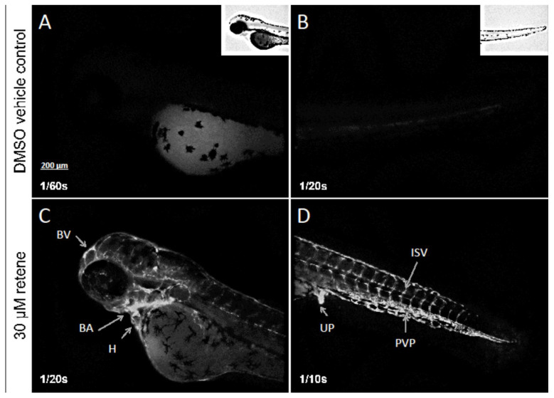
Figure 2
Whole-mount immunolocalization of zebrafish CYP1A protein in a representative DMSO vehicle (control) larva (
Acknowledgments
This image is the copyrighted work of the attributed author or publisher, and
ZFIN has permission only to display this image to its users.
Additional permissions should be obtained from the applicable author or publisher of the image.
Full text @ Toxics