IMAGE

Fig. 6

ID
ZDB-IMAGE-220725-6
Source
Figures for Ciampi et al., 2022
Image
Figure Caption

Fig. 6

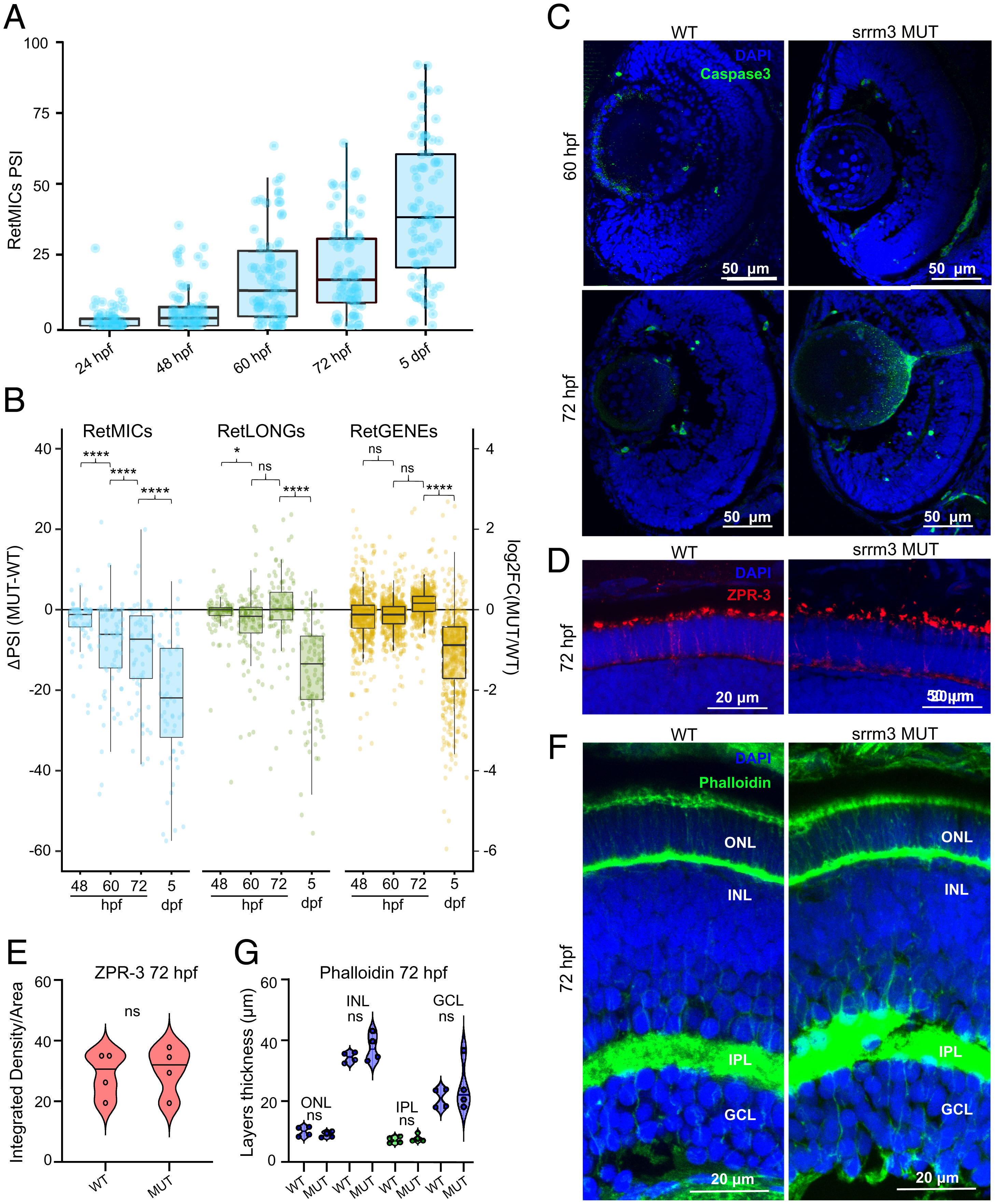
Transcriptomic and morphological alterations of srrm3 mutants throughout development. (A) Inclusion levels (PSIs) of RetMICs in WT zebrafish heads at 24 hpf and eyes at 48 hpf, 60 hpf, 72 hpf, and 5 dpf. (B) Box plots showing the distribution of changes in inclusion levels for RetMICs and RetLONGs [ΔPSI (MUT-WT)] (left y axis) and in expression levels for RetGENEs [log2FC(MUT/WT)] (right y axis) in WT and srrm3 MUT eyes at different time points. Only exons with sufficient read coverage across all conditions are shown. P values from paired one-tailed Wilcoxon tests. (C) Caspase3 staining for WT and srrm3 MUT retinae at 60 hpf and 72 hpf; n = 4 for all genotypes and conditions. (D and E) ZPR-3 staining (D) and positive area quantification (E) for WT and srrm3 MUT retinae at 72 hpf; n = 4. P values from unpaired t tests. (F and G) Phalloidin staining (F) and thickness analysis for different retinal layers (G) for WT and srrm3 MUT retinae. n = 4. P values from unpaired t tests.

Figure Data
Acknowledgments
This image is the copyrighted work of the attributed author or publisher, and ZFIN has permission only to display this image to its users. Additional permissions should be obtained from the applicable author or publisher of the image. Full text @ Proc. Natl. Acad. Sci. USA