
Fig. 2
- ID
- ZDB-IMAGE-220520-18
- Publication
- Pan et al., 2022 - Gnetum montanum extract induces apoptosis by inhibiting the activation of AKT in SW480 human colon cancer cells
- All Figures
- Figures for Pan et al., 2022
|
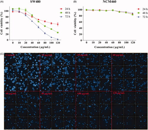
Fig. 2 Figure 2. Effect of GME on the viability of colon cancer cells SW480. (A) Colon cancer cells SW480 were treated with GME at concentrations of 0–120 μg/mL in triplicates for 24, 48, and 72 h. (B) The human normal colonic epithelial cells NCM460 were treated with GME at concentrations of 0–120 μg/mL in triplicates for 24, 48, and 72 h. Cell viability was measured by MTS assay. (C) The SW480 cells were treated with GME for 48 h. Apoptotic bodies were stained with DAPI (4′,6-diamidino-2-phenylindole). The cell morphology changed significantly, and the nucleus contracted and rounded. The cells were observed under the Operetta CLS high-content analysis system at DAPI pathway.