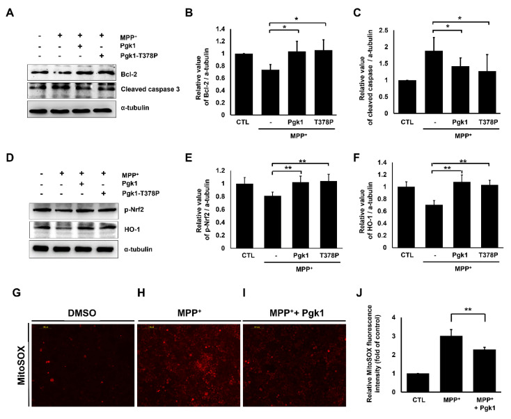
IMAGE

Figure 7

ID
ZDB-IMAGE-220430-92
Source
Figures for Lin et al., 2022
Image
Figure Caption

Figure 7

Extracellular addition of Pgk1 suppresses MPP+-induced apoptosis and mitochondrial ROS levels in SH-SY5Y cells. (A,D) Western blot analysis. The expression levels of Bcl-2, cleaved caspase-3, and p-Nrf2 and HO-1 proteins, as indicated, of SH-SY5Y cells treated or not treated with 0.5 mM MPP+ in the absence or presence of Pgk1 or kinase-deficient mutant T378P Pgk1 for 24 h. The α-tubulin served as an internal control of loading protein. (B,C,E,F) Statistical analysis of relative value of target protein expressed in cells with different treatment after normalization of the expression level of α-tubulin. (GI) The microscopic images of MitoSOX red fluorescence signal shown on cells. SH-SY5Y cells treated with or without 0.5 mM MPP+ in the absence or presence of Pgk1 and then stained with MitoSOX to detect the level of ROS generated in cells. (J) Quantification of intensity of fold increase of MitoSOX red fluorescence signal compared to that of cells without treatment served as a control group (CTL), which was set as 1. All data were averaged from three independent experiments and represented as mean ± S.D. Student’s t-test was used to determine significant differences between each group (**, p < 0.01; *, p < 0.05).

Acknowledgments
This image is the copyrighted work of the attributed author or publisher, and ZFIN has permission only to display this image to its users. Additional permissions should be obtained from the applicable author or publisher of the image. Full text @ Int. J. Mol. Sci.