
Image
|
Figure Caption
Figure 5
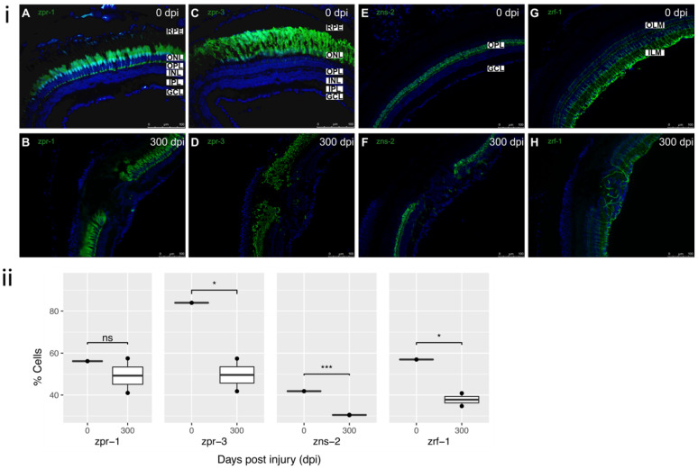
Figure 5. Cellular components of defect at 300 dpi: (i) (A) Uninjured retina (0 dpi) showing typical staining of cones with zpr-1 antibody. (B) Absence of restoration of cone photoreceptors within the injured retina at 300 dpi. (C) Uninjured retina (0 dpi) showing typical staining of rods with zpr-3 antibody. (D) Incomplete restoration and disorganization of rod photoreceptors within the lesion at 300 dpi. (E) Uninjured retina (0 dpi) showing typical staining of horizontal cells with zns-2 antibody. (F) Absence of horizontal cells within the lesion at 300 dpi. (G) Uninjured retina (0 dpi) showing typical staining of MG with zrf-1 antibody. ILM and OLM labelled for reference. (H) Partial restoration of MG within the residual lesion at 300 dpi. All sections were counterstained with DAPI. zpr-1, zpr-3, zns-2, zrf-1 markers—green; Nuclei, DAPI—blue. Retinal layers labelled in control panel A for reference; RPE—retinal pigment epithelium, OLM—outer limiting membrane; ONL—outer nuclear layer, OPL—outer plexiform layer, INL—inner nuclear layer, IPL—inner plexiform layer, GCL—ganglion cell layer; ILM—inner limiting membrane. Images (A,C) at 40×, rest of images at 20×. Scale bars represent 100 µm. (ii) Relative percentage of cell markers (zpr-1, zpr-3, zns-2, zrf-1) at 0–300 dpi (n = 2), * p value < 0.05; *** p value <0.005.
Acknowledgments
This image is the copyrighted work of the attributed author or publisher, and
ZFIN has permission only to display this image to its users.
Additional permissions should be obtained from the applicable author or publisher of the image.
Full text @ Cells