
Image
|
Figure Caption
Figure 3
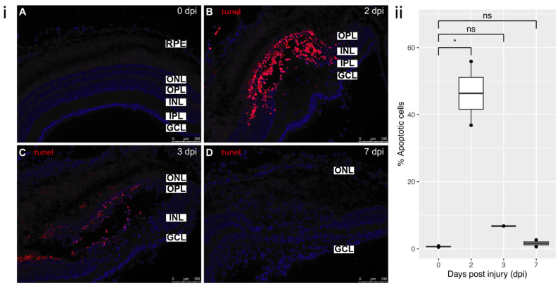
Figure 3. TUNEL staining: (i) (A) Control staining in uninjured retina (0 dpi). (B) 2 dpi: profound localized cell death in the area of cryoinjury, degeneration phase. Predominantly affected layers were OPL and INL. (C) 3 dpi: cell death still ongoing, but to a lesser extent than at 2 dpi, clearing off the dead cells by macrophages. (D) 7 dpi: damaged nuclei are no longer detected in the retina, reprogramming of MG, remodeling phase. All sections were counterstained with DAPI. Apoptotic cells—TMR Red; Nuclei, DAPI—blue. Retinal layers labelled in control panel for reference; RPE—retinal pigment epithelium, ONL—outer nuclear layer, OPL—outer plexiform layer, INL—inner nuclear layer, IPL—inner plexiform layer, GCL—ganglion cell layer. Scale bars represent 100 µm. (ii) Relative percentage of apoptotic cells at each time-point (n = 2), * p value = 0.02.
Acknowledgments
This image is the copyrighted work of the attributed author or publisher, and
ZFIN has permission only to display this image to its users.
Additional permissions should be obtained from the applicable author or publisher of the image.
Full text @ Cells