IMAGE

Figure 3.

ID
ZDB-IMAGE-220411-7
Source
Figures for Sheng et al., 2022
Image
Figure Caption

Figure 3.

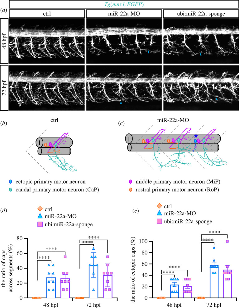
Deficiency of mir-22a caused aberrant axonal projection of PMNs. (a) Confocal imaging analysis of primary motor neurons in the control embryos, miR-22a morphants and Tg(mnx1:GFP::ubi:miR-22a-sponge) embryos at 48 and 72 hpf. Arrowheads indicate the CaPs across different segments. (b,c) Schematic diagram for primary motor neurons in the control embryos and miR-22a morphants. (d) Statistical analysis of the ratio of CaPs across different segments in the control (n = 8), miR-22a morphants (n = 8) and Tg(mnx1:GFP::ubi:miR-22a-sponge) embryos (n = 8) at 48 and 72 hpf; One-way ANOVA, ****p < 0.0001. (e) Statistical analysis of the ratio of aberrant axonal projection of CaPs in the control (n = 8), miR-22a morphants (n = 8) and Tg(mnx1:GFP::ubi:miR-22a-sponge) embryos (n = 8) at 48 and 72 hpf; one-way ANOVA, ****p < 0.0001.

Acknowledgments
This image is the copyrighted work of the attributed author or publisher, and ZFIN has permission only to display this image to its users. Additional permissions should be obtained from the applicable author or publisher of the image. Full text @ Open Biol.