IMAGE

Figure 1.

ID
ZDB-IMAGE-220411-5
Source
Figures for Sheng et al., 2022
Image
Figure Caption

Figure 1.

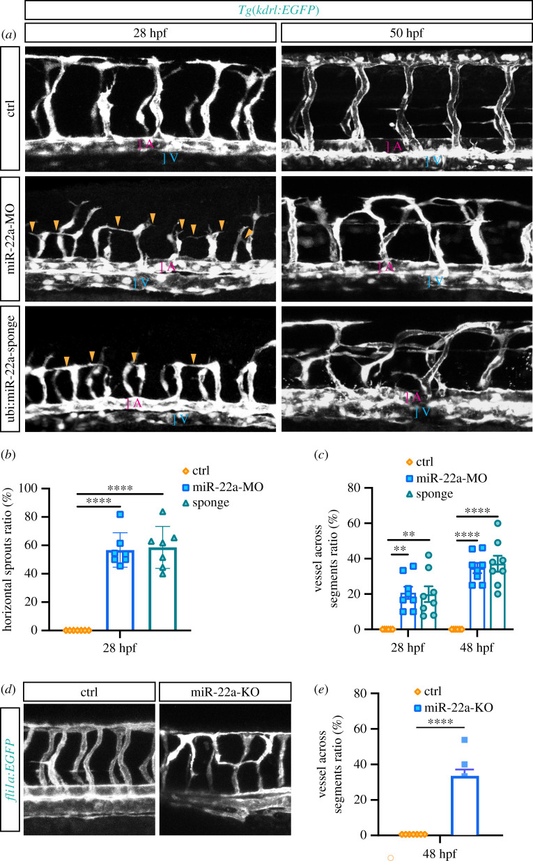
Deficiency of miR-22a caused aberrant vascular networks. (a) Confocal imaging analysis of control MO, miR-22a-MO and miR-22a-sponge-injected Tg(kdrl:EGFP) embryos at 30 hpf and 48 hpf. The arrowheads indicate the aberrant angiogenic sprouts. A: dorsal aorta; V: posterior cardinal vein. (b) Statistics of horizontal sprouts ratio in 30 hpf embryos injected with control MO (n = 7), miR-22-MO (n = 7) and miR-22-sponge (n = 7). One-way ANOVA, **** p < 0.0001. (c) Statistics of embryos with vessel across segments ratio in each group: control MO (n = 6), miR-22a-MO (n = 8) and miR-22-sponge (n = 8). One-way ANOVA, ** p < 0.01; **** p < 0.0001. (d) Confocal imaging analysis of control and miR-22a-KO Tg(fli1a:EGFP-CAAX) embryos at 48 hpf. (e) The statistics of embryos with vessels across segments ratio in each group: control (n = 7) and miR-22-KO (n = 7). t-test, ****, p < 0.0001.

Acknowledgments
This image is the copyrighted work of the attributed author or publisher, and ZFIN has permission only to display this image to its users. Additional permissions should be obtained from the applicable author or publisher of the image. Full text @ Open Biol.