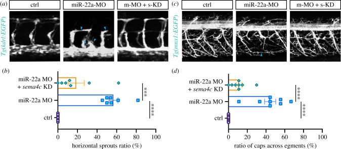
IMAGE

Figure 6.

ID
ZDB-IMAGE-220411-10
Source
Figures for Sheng et al., 2022
Image
Figure Caption

Figure 6.

Reducing sema4c partially restored the defects of ISVs and PMNs in miR-22a-deficient embryos. (a) Confocal imaging analysis of blood vessels in control, miR-22a-MO, and miR-22a-MO + sema4c knockdown embryos at 30 hpf. (b) Statistics of horizontal sprouts ratio in control (n = 7), miR-22a-MO (n = 7) and miR-22a-MO + sema4c knockdown embryos (n = 7) at 30 hpf. One-way ANOVA; ***p < 0.001; ****p < 0.0001. (c) Confocal imaging analysis of control, miR-22a-MO and miR-22a-MO + sema4c knockdown Tg(mnx1:EGFP) embryos at 72 hpf. (d) The statistical analysis of the ratio of aberrant axonal projection of CaPs in the control (n = 8), miR-22a morphants (n = 8) and miR-22a-MO + sema4c knockdown embryos (n = 8) at 72 hpf; one-way ANOVA, ****p < 0.0001.

Acknowledgments
This image is the copyrighted work of the attributed author or publisher, and ZFIN has permission only to display this image to its users. Additional permissions should be obtained from the applicable author or publisher of the image. Full text @ Open Biol.