
|
Figure 1
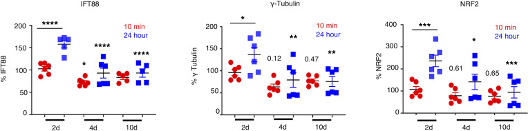
Shear stress causes brain ECs to express fewer cilia proteins in vitro. HBMVECs were subjected to graded strengths of shear stress (2 dyne/cm2, 4 dyne/cm2, and 10 dyne/cm2) by the Ibidi flow system. A total of 2 dyne/cm2 was utilized as a “steady-state” flow condition. Following flow with durations as indicated, the expression of cilia-associated proteins was quantified as MFI by flow cytometry. NRF2, the transcription factor reported to control cilia formation and function, was also included in the study. Expression of proteins in the samples was normalized against their respective “no flow” controls. ANOVA was performed to compare between the experimental groups versus steady-state control group for 10-minute or 24-hour time points. ANOVAs were 2 way. Analysis was also performed between 10 minutes and 24 hours in 2 dyne/cm2 group. In all 3 protein expressions, no statistical difference was observed between 4 dyne/cm2 and 10 dyne/cm2 groups. *P < 0.05, **P < 0.01, ***P < 0.001, ****P < 0.0001. For all groups reported in this figure, n = 6 except for IFT88 (n = 5 for 10 dyne group).