Image
Figure Caption

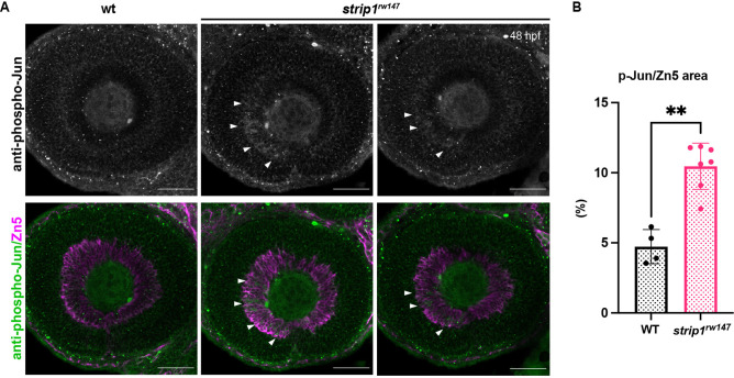
Figure 6—figure supplement 2—source data 1. Jun is activated in retinal ganglion cells (RGCs) of <italic toggle='yes'>strip1</italic> mutants at 48 hpf.

(A) Whole-mount labeling of 48-hpf wild-type and strip1rw147 mutant retinas with anti-phospho-Jun (p-Jun) antibody and zn5 antibody, which label active Jun and RGCs, respectively. Arrowheads show RGCs expressing phospho-Jun in the ventronasal region. Scale bar, 50 μm. (B) Percentage of phospho-Jun area relative to zn5 area at 48 hpf. Mann–Whitney U-test, n ≥ 4. Data are represented as means ± standard deviation (SD). **p < 0.01.

Data for <xref rid='fig6s2' ref-type='fig'>Figure 6—figure supplement 2B</xref>.

Acknowledgments
This image is the copyrighted work of the attributed author or publisher, and ZFIN has permission only to display this image to its users. Additional permissions should be obtained from the applicable author or publisher of the image. Full text @ Elife