IMAGE

Fig 7

ID
ZDB-IMAGE-220318-8
Genes
Source
Figures for Nandamuri et al., 2022
Image
Figure Caption

Fig 7 <italic toggle='yes'>dzip1</italic> mutants exhibit defective accumulation of POM-derived endothelial cells.

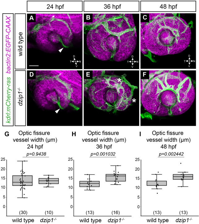
(A-F) Embryos visualized for endothelial cells (green; Tg(kdrl:ras-mCherry)) and cell membranes (magenta; Tg(bactin2:EGFP-CAAX)) in wild type (A-C) and dzip1ts294e mutants (D-F) at 24 hpf (A, D), 36 hpf (B, E), and 48 hpf (C, F). All images are lateral views of 3-dimensional renderings. mCherry-positive endothelial cells populate the optic fissure by 24 hpf in both wild type and dzip1-/- embryos (A, D; white arrowheads). (E) asterisks, ectopic branching of the superficial network in dzip1 mutant. (G-I) Quantification of fissure vessel width (in μm) at 24 hpf, 36 hpf, and 48 hpf. n (embryos) for each genotype shown at the base of the graph. P-values were calculated using Welch’s t-test (M-O). Scale bar: 50 μm. D, dorsal; V, ventral; N, nasal; T, temporal.

Figure Data
Acknowledgments
This image is the copyrighted work of the attributed author or publisher, and ZFIN has permission only to display this image to its users. Additional permissions should be obtained from the applicable author or publisher of the image. Full text @ PLoS One