IMAGE

Fig. 3.

ID
ZDB-IMAGE-220317-10
Genes
Source
Figures for Serrano et al., 2021
Image
Figure Caption

Fig. 3.

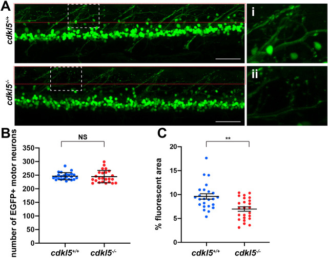
Assessment of neuronal pathology in cdkl5−/− fish. (A) Maximum intensity projection confocal images of EGFP-labelled motor neurons from Tg(islet1:EGFP); cdkl5+/+ (white dashed box zoomed in i) and Tg(islet1:EGFP); cdkl5−/− (white dashed box zoomed in ii) along the spinal region at 6 dpf. Red dashed boxes indicate the regions quantified in C. Scale bars: 50 µm. (B) Quantification of the number of EGFP+ motor neurons at 6 dpf. Data are mean±95% c.i. for three independent experiments [n=8, 8 and 8 for Tg(islet1:EGFP); cdkl5+/+, and n=8, 8 and 8 for Tg(islet1:EGFP); cdkl5−/− fish]. (C) Quantification of the mean percentage area of fluorescence from Tg(islet1:EGFP); cdkl5+/+ and Tg(islet1:EGFP); cdkl5−/− (within the red dashed box region in A) at 6 dpf. Data are mean±s.e.m. for three independent experiments [n=8, 8 and 8 for Tg(islet1:EGFP); cdkl5+/+, and n=8, 8 and 8 for Tg(islet1: EGFP); cdkl5−/− fish]. **P<0.01; NS, not significant (two-tailed t-test).

Figure Data
Acknowledgments
This image is the copyrighted work of the attributed author or publisher, and ZFIN has permission only to display this image to its users. Additional permissions should be obtained from the applicable author or publisher of the image. Full text @ Dis. Model. Mech.