IMAGE

Figure 4

ID
ZDB-IMAGE-220303-14
Source
Figures for Benardais et al., 2022
Image
Figure Caption

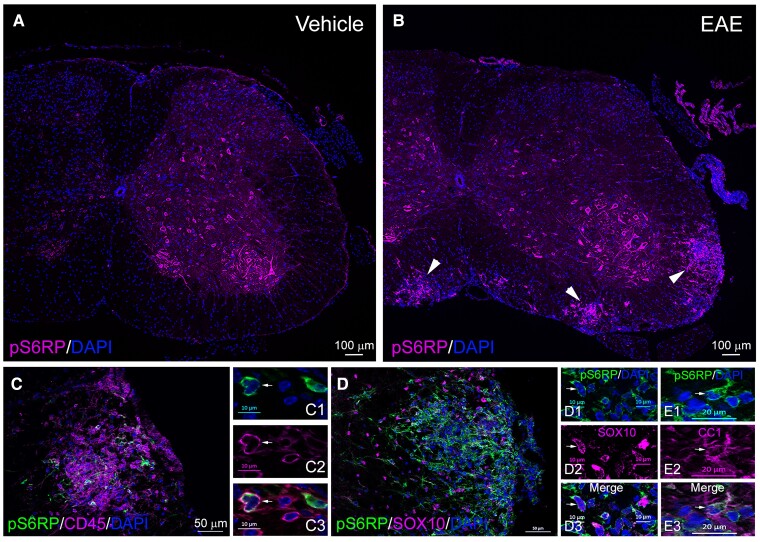
Figure 4

pS6RP expression during EAE in mouse spinal cord. (A) pS6RP immunolabelling in the naive adult spinal cord. Note that pS6RP is mainly detected in motor neurons and absent in white matter in the naive adult spinal cord. (B) pS6RP immunostaining in MOG-induced EAE lesions of the spinal cord. Inflammatory foci are indicated by arrowheads. (CE3) pS6RP-expressing cells within EAE lesions are predominantly CD45+ cells (C, insets C1–C3) or Sox10+ (D, insets D1–D3) and CC1+ (insets E1–E3) oligodendrocytes. Arrows in insets indicate double-positive cells. Scale bars: (A and B), 100 µm; (C and D), 50 µm; (C1–C3, D1–D3), 10 µm; (E1–E3), 20 µm.

Acknowledgments
This image is the copyrighted work of the attributed author or publisher, and ZFIN has permission only to display this image to its users. Additional permissions should be obtained from the applicable author or publisher of the image. Full text @ Brain Commun