
|
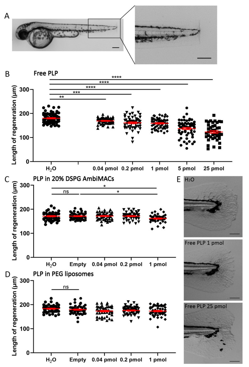
Figure 5
Liposome encapsulation of PLP decreases its inhibitory effect on regeneration of the tail fin after amputation. Embryos (at 2 dpf) were subjected to the tail fin amputation procedure and injected with different doses of free or liposome-encapsulated PLP. (A) Representative image of a 2 dpf zebrafish embryo immediately after amputation, showing the position of the amputated part of the tail fin. Scale bar = 200 μm. (B–D) The length of the regenerated tail fin, measured at 36 h post-amputation (hpa), are shown after injection of different doses of free PLP (B), PLP encapsulated in AmbiMACs (20% DSPG) (C) and PLP encapsulated in PEGylated liposomes (D); H2O and empty liposomes were injected as control. Statistical analysis was performed by one-way ANOVA with Bonferroni’s post hoc test. Significant inhibition of the tail fin regeneration was observed when embryos had been injected with 0.04–25 pmol of free PLP or 1 pmol of PLP encapsulated in AmbiMACs (20% DSPG). No significant inhibition was observed after injection with PLP encapsulated in PEGylated liposomes. Each data point represents a single embryo, and the means ± SEM of the data accumulated from three independent experiments are shown in red. Statistically significant differences between groups are indicated by: ns, non-significant; * p < 0.05; ** p < 0.01; *** p < 0.001; **** p < 0.0001. (E) Representative images of regenerated tail fins at 36 hpa for embryos injected with H2O, 1 pmol PLP, or 25 pmol PLP. Scale bar = 100 μm.