
|
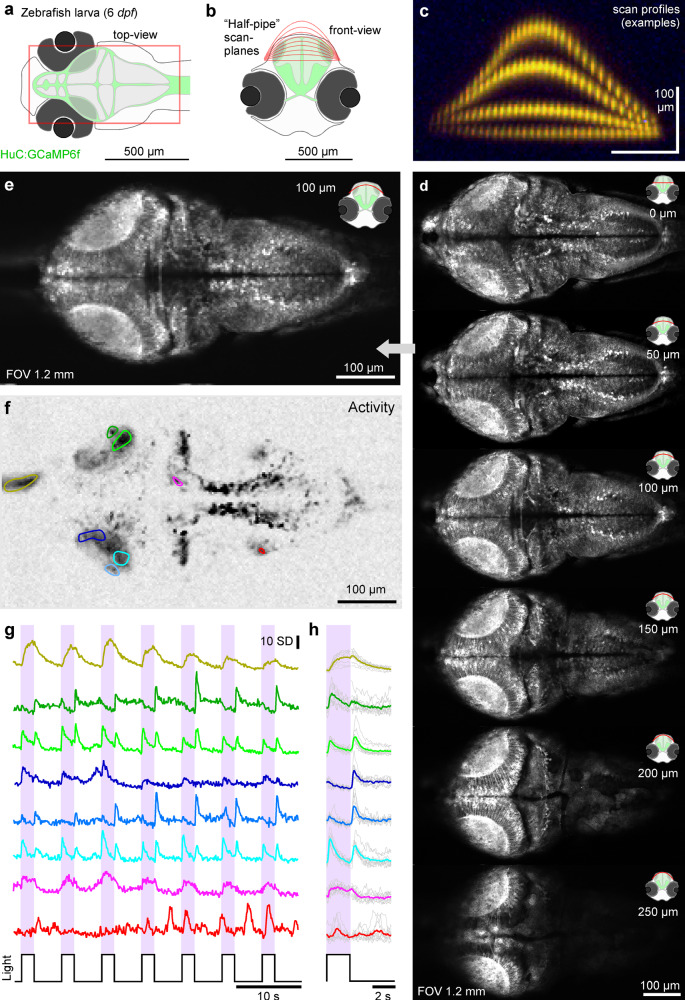
Fig. 7
a–c Schematic of HuC:GCaMP6f larval zebrafish brain viewed from top (a) and front (b) with scan planes indicated, and (c) example-scan-profiles. d nTC1 512 × 1024 scans of a 6 dpf zebrafish brain with different plane curvatures, with peak axial displacement at scan centre as indicated. At curvatures ~100–150 µm peak displacement the scan approximately traverses the surface of the tectum. e–h Mean (e), activity-correlation (f, cf. Fig. 3c) and fluorescence traces (g, raw and h, event-triggered mean) from a 170 × 340 px scan (5.88 Hz) of the 100 µm peak displacement configuration (image 3 in (d)). The fish was presented with full-field and spectrally broad (~360–650 nm) series of light-flashes. See also Fig. S4.