
|
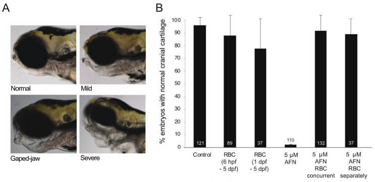
Figure 1 AFN causes craniofacial defects which are rescued by RBC. (A) Representative images of larvae at 5 dpf (lateral view) with normal cartilage (top left) and abnormal cartilage; mild (top right), gaped jaw (bottom left) and severe (bottom right) as a result of 5 μM AFN treatment. (B) Proportion of larvae with normal craniofacial cartilage in untreated controls, RBC (500 µM) exposure 6 hpf–5 dpf, RBC (500 µM) exposure 1–5 dpf, 5 µM AFN only, 5 µM AFN with concurrent exposure to RBC (500 µM) starting at 6 hpf through to 5 dpf, 5 µM AFN with separate exposure to RBC (500 µM) with RBC treatment commencing at 1 dpf through to 5 dpf. Error bars show the standard error of the mean from three biological replicates. Data labels show the total number of larvae observed for each treatment group. Auranofin, AFN; Riboceine, RBC; hours post fertilization, hpf; days post fertilization, dpf.