IMAGE

Fig. 1

ID
ZDB-IMAGE-220131-223
Source
Figures for Liu et al., 2021
Image
Figure Caption

Fig. 1

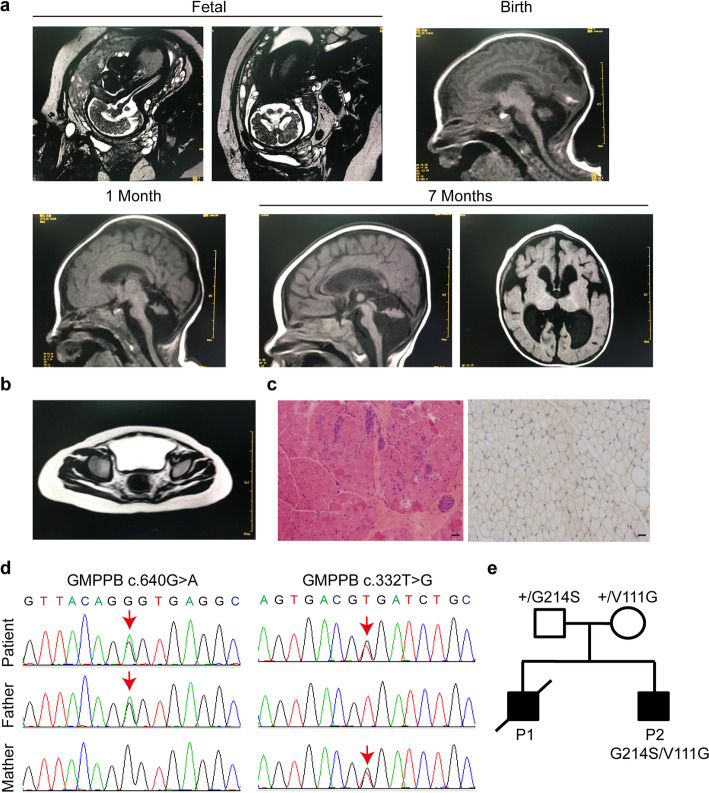
Clinical and genetic characterization. a Brain MRI of the patient at fetal 34 weeks, birth, 1 month, and 7 months, revealing evidence of cerebellar hypoplasia. b Muscle MRI in the right gluteus maximus muscle at 14 months. c Muscle biopsy from gastrocnemius at 14 months. The diameter of muscle fibers range between 5 and 50 μm, with occasional degenerated and regenerated muscle fibers. Scale bar: 50 μm d Sequence chromatograms of GMPPB gene of the patient. Variations and corresponding WT sequences are shown. The patient carries two missense mutations of GMPPB gene (arrows), inherited from his mother (c.332 T > G) and father (c.640G > A), respectively. e Pedigrees

Acknowledgments
This image is the copyrighted work of the attributed author or publisher, and ZFIN has permission only to display this image to its users. Additional permissions should be obtained from the applicable author or publisher of the image. Full text @ Mol Biomed