
Fig. 5
- ID
- ZDB-IMAGE-220107-5
- Publication
- Graves et al., 2021 - Zebrafish harbor diverse intestinal macrophage populations including a subset intimately associated with enteric neural processes
- All Figures
- Figures for Graves et al., 2021
|
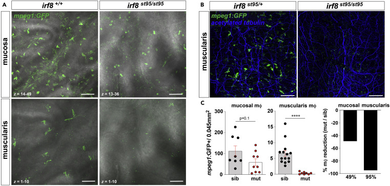
Fig. 5 (A) Representative confocal z stack micrographs of ex vivo irf8+/+ and irf8−/− Tg(mpeg1:GFP) proximal gut tissue showing irf8-mediated mφ loss in both the mucosa (top) and muscularis (bottom) (40 X,1 μm voxel depth). (B) Representative confocal z stack micrographs of ex vivo irf8+/+ and irf8−/− Tg(mpeg1:GFP) proximal gut-fixed tissue (anti-acetylated tubulin antibody retrieval) showing irf8-mediated neural process-associated MMφ loss in the muscularis (20 X, z = 16, 2 μm voxel depth). (C) Quantification of mpeg1:GFP + cells in the mucosa (left) and muscularis (middle) of irf8st95/st95 mutants (mut) compared to wild-type or heterozygous siblings (sib) showing ~50% reduction in mucosal macrophages versus a 95% reduction of muscularis macrophages (right) from pooled non-overlapping confocal z stack micrographs (n = 3–4 individuals per group). More inter-individual variation was observed in frequency of mucosal macrophages which in part may be due to influence of the gut microbiota. Data shown are mean ± S.E.M. ∗∗∗∗p < 0.0001 determined by nonparametric Mann-Whitney two-tailed T test. Data are representative of at least n = 3 adult individuals per group and at least 3 independent experiments. Scale bar = 100 μm.