
|
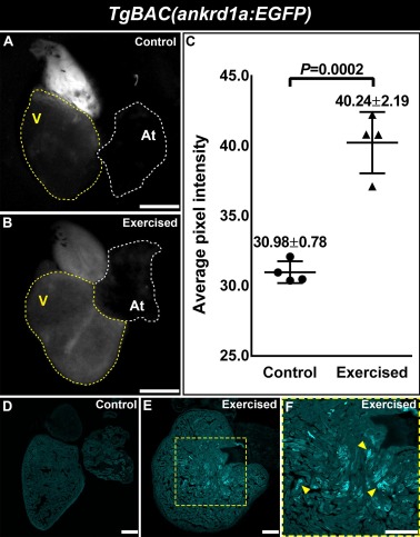
Fig. 3 Exercise upregulates TgBAC(ankrd1a:EGFP) expression in the heart of adult zebrafish. (A, B) Fluorescence images of control and exercised hearts. Atrial (At) and ventricular (V) chambers are delineated by white and yellow dotted lines, respectively. (C) EGFP signal was quantified in control and exercised hearts (n = 4) and presented as average pixel intensity ± SD. (D, E) Representative images of cryosections of control and exercised hearts. (F) Magnified area from E, yellow arrows point to cells with pronounced transgene expression. Scale bars, 500 µm for A and B, 100 µm for D to F. (For interpretation of the references to color in this figure legend, the reader is referred to the web version of this article.)
Reprinted from Gene, 792, Boskovic, S., Marin Juez, R., Stamenkovic, N., Radojkovic, D., Yr Stainier, D., Kojic, S., The stress responsive gene ankrd1a is dynamically regulated during skeletal muscle development and upregulated following cardiac injury in border zone cardiomyocytes in adult zebrafish, 145725, Copyright (2021) with permission from Elsevier. Full text @ Gene