
|
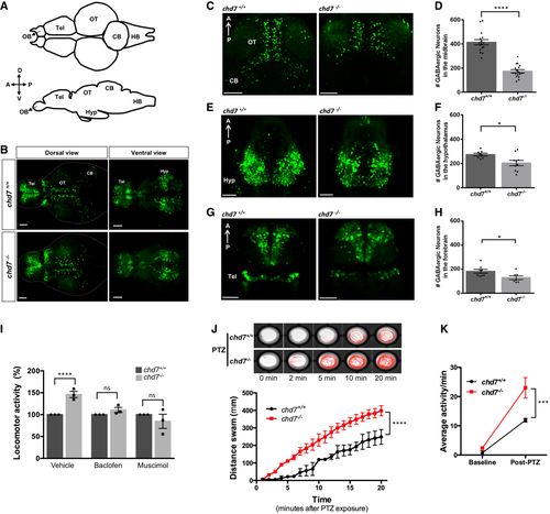
Fig. 2 A. Structural illustration of 5 dpf zebrafish brain from dorsal (top) and lateral (bottom) view (OB: Olfactory bulb, Tel: Telencephalon, OT: Optic tectum, CB: Cerebellum, HB: Hindbrain). B. 5 dpf dlx5a/6a transgenic line showing GFP+ GABAergic neurons are reduced in chd7−/− mutants (bottom) in comparison with controls (top) in both dorsal (left) and ventral (right) view. C–H. Total number of GABAergic neurons (GFP+ cells) in (C, D) the optic tectum (OT) and cerebellum (CB) regions of 5 dpf wild-type and chd7 mutant fish (n = 16; ****P < 0.0001; Student’s unpaired t-test), (E, F) the hypothalamus (hyp) region (n = 10; *P = 0.0182; Student’s t-test) and (G, H) the telencephalon (tel) (n = 7; *P = 0.0347; Student’s t-test). I. Treatment of control (dark grey) and mutants (light grey) with GABA agonists Baclofen (N = 3, n = 24; ns, P = 0.1427; Student’s t-test) and Muscimol (N = 3, n = 24; ns, P = 0.3987; Student’s t-test) showing recovery of hyperactive locomotor activity in chd7−/− mutants (vehicle: N = 3, n = 24, ****P < 0.0001; Student’s t-test). J. Functional analysis of GABAergic signalling shows increased responsiveness to GABA antagonist PTZ in both onset and overall locomotor activity (n = 24; ****P < 0.0001; one-way ANOVA). K. Average locomotor activity between 2 dpf controls (black) and chd7−/− mutants (red) shows increased activity after 3 mM PTZ exposure (n = 24; ***P < 0.001; two-way ANOVA). Data information: Data are presented as mean ± SEM. Scale bar = 50 μm. n represents number of fish used. N represents number of experimental repeats.