
|
Fig. 4
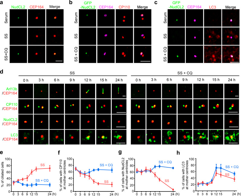
a MEF cells were cultured with or without serum starvation, treated with or without 20 μM CQ, and subjected to immunofluorescence analysis with anti-NudCL2 and anti-CEP164 antibodies. b MEF cells were transfected with GFP-NudCL2 and applied for immunofluorescence analysis with anti-CP110 and anti-CEP164 antibodies under the indicated treatments. c MEF cells transfected with GFP-NudCL2 were stained with anti-LC3 and anti-CEP164 antibodies under the indicated treatments. d–h MEF cells treated with or without 20 μM CQ were starved for different times, and then subjected to immunostaining analyses with the indicated antibodies. The percentages of ciliated cells (e), cells with CP110 dot at mother centrioles (f), cells with NudCL2 dot at mother centrioles (g), and cells with LC3-positive puncta at mother centrioles (h) are shown. Scale bars, 2 µm.