
|
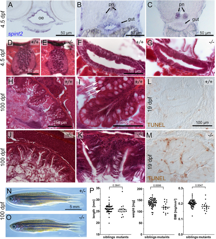
Fig. 2 A-C. Cross sections of a 4.5 dpf embryo following spint2 WISH. spint2 expression is strongly detected in the oral epithelium (oe), moderately in the anterior gut (B), and decreasing towards more posterior regions, in contrast to strong spint2 signal in the pronephric ducts (pn) (C). D-G. H&E staining of cross sections of 4.5 dpf spint2 siblings (D, F) and mutants (E, G) showing normal spinal cord morphology (D, E) and gut epithelium (F, G). H–K. H&E staining of cross sections of the rostral intestine of an adult (100 dpf) wild-type sibling (H–I) and a spint2 mutant (J-K). The mutant intestine contains fewer goblet cells (arrows) and a disrupted epithelial integrity. L,M. TUNEL staining of cross sections of the rostral intestine of an 19 dpf juvenile wild-type sibling (L) and a spint2 mutant (M). N,O. Images of an adult (100 dpf) wild-type sibling (N) and a spint2 mutant (O) male raised together. The mutant is of slightly smaller size. P. Quantification of length in mm, weight in mg, and BMI in mg/mm2 of spint2 sibling and mutant males raised together. Dots indicate individual fish, lines represent the mean, and p values were determined by a Student’s t-test. Four experiments/families; 72 individuals.
Reprinted from Developmental Biology, 476, Hatzold, J., Wessendorf, H., Pogoda, H.M., Bloch, W., Hammerschmidt, M., The Kunitz-type serine protease inhibitor Spint2 is required for cellular cohesion, coordinated cell migration and cell survival during zebrafish hatching gland development, 148-170, Copyright (2021) with permission from Elsevier. Full text @ Dev. Biol.