
|
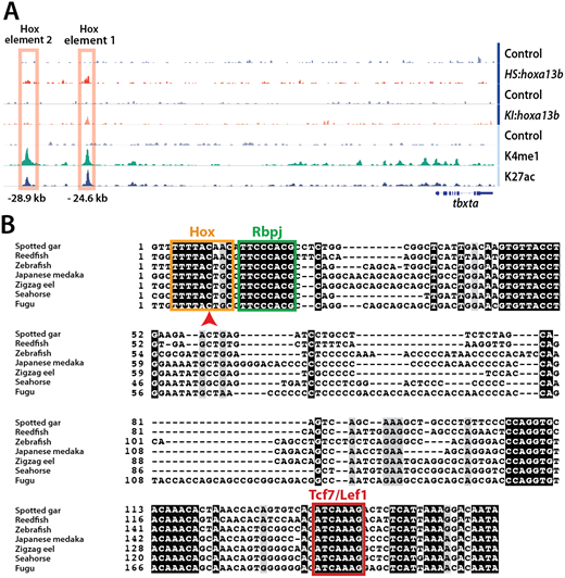
Fig. 4 Hox elements upstream of the tbxta transcriptional start site. (A) IGV tracks showing the two Hoxa13b-binding elements discovered using CUT&RUN. A peak appears at the Hox element 1 site in both the HS:hoxa13b-FLAG-GFP and KI:hoxa13b-FLAG-GFP lines, whereas a weaker signal exists at the Hox element 2 site in the HS:hoxa13b-FLAG-GFP sample but not in the KI:hoxa13b-FLAG-GFP sample. Both elements have peaks for H3K4me1 and H3K27ac. (B) The alignment of Hox element 1 in a variety of species is shown, including consensus sites for Hox, Rbpj and Tcf7/Lef1 binding. In all species, there is a C in the Hox site at the nucleotide position specifically recognized by posterior Hox proteins (arrowhead).