
|
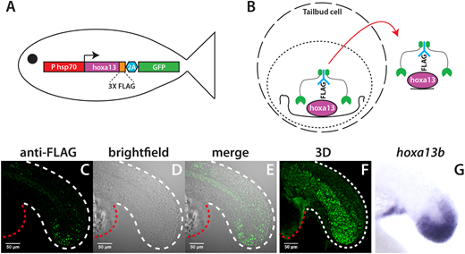
Fig. 1 Identification of in vivo Hoxa13b targets with CUT&RUN. (A) Schematic of the HS:hoxa13b-FLAG-GFP transgenic line. (B) CUT&RUN procedure. Cells isolated from dissected tailbuds are separated and permeabilized. First, an antibody is added (in this case anti-FLAG), then a Protein A/G-Micrococcal nuclease (green), which cuts the genomic DNA locally, allowing transcription factor-DNA complexes to exit the cell, whereupon they are isolated and analyzed by DNA sequencing. (C-F) Expression of Hoxa13b-FLAG in KI:hoxa13b-FLAG-GFP embryos as detected with an anti-FLAG antibody in a 20-somite stage embryo. Shown are the lateral view of a middle slice of the tail (C-E) and an image from the 3D reconstruction (F). The white dashed line shows the posterior body border and the red dashed line shows the yolk extension. See also Movie 1 for a 360° view of the tail. (G) In situ hybridization showing hoxa13b transcripts in the tailbud of a 20-somite stage WT embryo for comparison.