IMAGE

Figure 5

ID
ZDB-IMAGE-211029-105
Source
Figures for D'Gama et al., 2021
Image
Figure Caption

Figure 5

foxj1a and foxj1b play diverse roles in the formation of motile ciliated cells

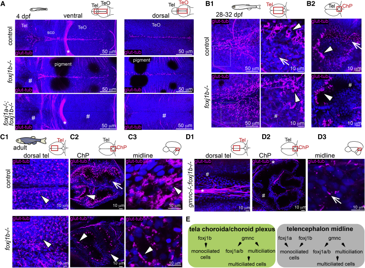
(A) Lack of cilia in the dorsal telencephalon in foxj1b mutant (middle; n = 9) and throughout the larval ventricular system in double foxj1a/b mutant (n = 6) at 4 dpf. Maximum projection at two different depths is shown.

(B1 and B2) MCCs (arrowhead) are not affected in the foxj1b mutant on the 1-month dorsal telencephalon (B1; n = 6) and ChP (B2; n = 6). Cilia remain absent in the monociliated cells (arrow) located laterally to the MCCs as shown in the magnified inset.

(C1–C3) MCCs are not affected in the adult medial TC (C1; n = 3), ChP (C2; n = 4), and midline of the telencephalon/diencephalon (C3; n = 3). (C2) Monociliated cells are particularly affected in the ChP.

(D1–D3) Loss of cilia in the medial TC (D1) and ChP (D2) of adult gmnc;foxj1b double mutants (n = 4). (D3) Single cilia remained in the telencephalic midline of double mutants (arrow) (n = 4).

(E) Diverse genetic programs instruct the formation of ciliated cells in the TC/ChP and in the telencephalon midline.

TeO, optic tectum; sco, sub-commissural organ. Arrows indicate monociliated cells, arrowheads indicate MCCs, and asterisks indicate axon tracts. Cilia loss is indicated by hashtag symbols.

See also Figures S4 and S5.

Acknowledgments
This image is the copyrighted work of the attributed author or publisher, and ZFIN has permission only to display this image to its users. Additional permissions should be obtained from the applicable author or publisher of the image. Full text @ Cell Rep.