IMAGE

Fig. 3

ID
ZDB-IMAGE-211025-59
Source
Figures for Brun et al., 2021
Image
Figure Caption

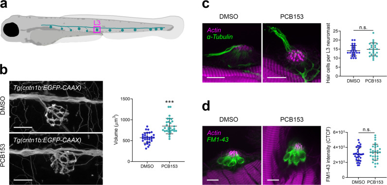
Fig. 3 Effect of PCB153 on morphology and function of lateral line hair cells and innervating neurons.

a Diagram indicating L3 neuromast. b Hair cell innervating neurons at neuromast L3 shown using Tg(cntn1b:EGFP-CAAX) and volume of hair cell innervating neurons of DMSO (n = 30) and PCB153 (n = 29) treated larvae. Scale bar = 20 µm. c Whole-mount immunostaining of kinocilia and hair cell innervating neurons (anti-acetylated tubulin antibody; green) and counterstain of stereocilia bundles and muscle tissue (actin; magenta) of DMSO (n = 26) and PCB153 (n = 24) treated larvae. The hair cell number was determined based on the number of kinocilia. Scale bar = 10 µm. d Uptake of FM1-43 dye as indication of functional hair cells of DMSO (n = 32) and PCB153 (n = 28) treated larvae. CTCF = Corrected total cell fluorescence. Scale bar = 10 µm. All data points are biologically independent samples from three independent experiments and mean ± SD are shown in the plots. Unpaired two-tailed t-test with Welch’s correction was used for statistical analysis in (b, c), a two-way ANOVA accounting for trial and treatment in (d). Asterisks indicate significant differences to controls (***p < 0.001).

Acknowledgments
This image is the copyrighted work of the attributed author or publisher, and ZFIN has permission only to display this image to its users. Additional permissions should be obtained from the applicable author or publisher of the image. Full text @ Commun Biol