
|
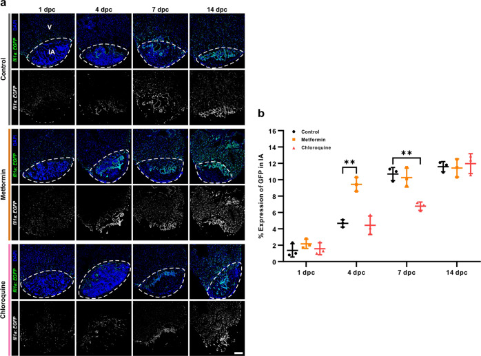
Fig. 4
a After heart cryoinjury, the Tg(fli1a:EGFP) fish, which express EGFP specifically in the endocardium and vascular endothelium, were maintained in untreated fish water (control) or in fish water containing 50 µM metformin or 100 µM CQ for 1–14 days. The hearts were then isolated, fixed, sectioned and immunostained with an anti-GFP antibody (in green), and co-labeled with DAPI to show the nuclei (in blue). V: ventricle; IA: injured area. Scale bar: 100 µm. b The percentage of Fli1a expressing cells in the injured area of the control, metformin and CQ treated fish at 1 dpc, 4 dpc, 7 dpc, and 14 dpc was quantified. The data are presented as mean ± SD, n = 3 hearts, **P < 0.01 vs control.