
Image
|
Figure Caption
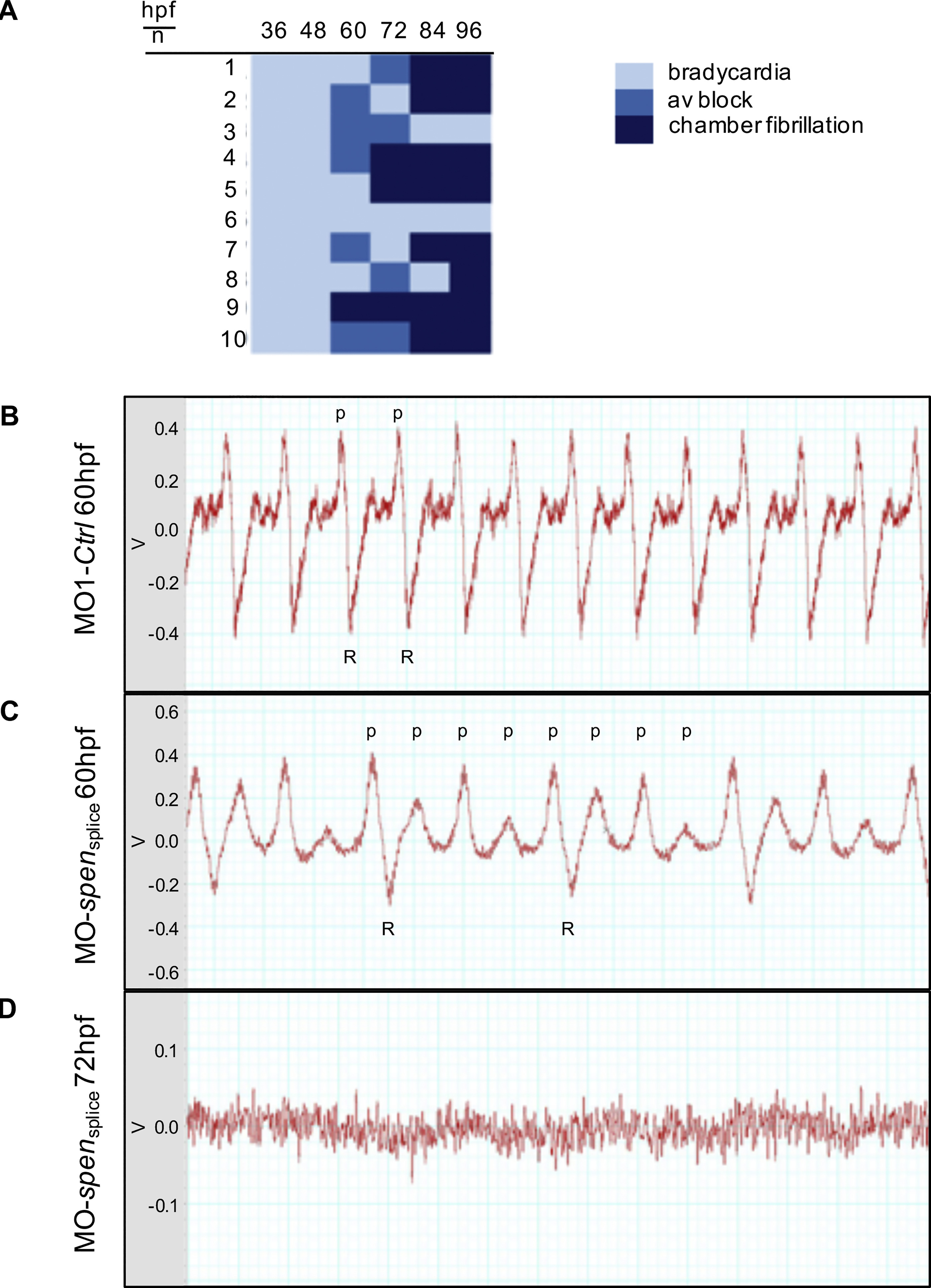
Fig. 3 (A) Overview of the heart rhythm phenotype of ten Spen-deficient embryos at different developmental time points. (hpf: hours post fertilization). (B-D) Zebrafish electrocardiograms. While in the controls every atrial excitation (p: p-wave) is followed by a ventricular potential (R: R-wave); (B), spen morphants show atrioventricular conduction abnormalities, as not every p-wave is followed by an R-wave (C). At 72 hpf only an uncoordinated atrial and ventricular electric activity can be documented, resembling atrial and ventricular fibrillation (D). V: Volt; Images (B,C,D) have been cropped.
Figure Data
Acknowledgments
This image is the copyrighted work of the attributed author or publisher, and
ZFIN has permission only to display this image to its users.
Additional permissions should be obtained from the applicable author or publisher of the image.
Reprinted from Journal of Molecular and Cellular Cardiology, 155, Rattka, M., Westphal, S., Gahr, B.M., Just, S., Rottbauer, W., Spen deficiency interferes with Connexin 43 expression and leads to heart failure in zebrafish, 25-35, Copyright (2021) with permission from Elsevier. Full text @ J. Mol. Cell. Cardiol.