IMAGE

Figure 5

ID
ZDB-IMAGE-211002-6
Source
Figures for Chebli et al., 2021
Image
Figure Caption

Figure 5

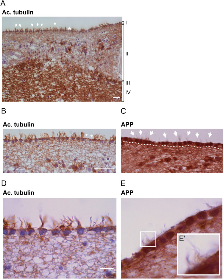
APP is localized to human ependymal cilia. (A) Brightfield overview of a human brain section of the caudate nucleus immunostained with an anti-acetylated tubulin antibody reveals the different cellular layers (I–IV): (I) ependymal layer with motile cilia orienting towards the ventricle lumen, (II) cellular extensions connecting the ependymal cells, (III) cellular layer dense in astrocytes, (IV) brain parenchyma. (BE) Higher magnifications of the ependymal layer show clear cilia (acetylated tubulin (B,D)) and APP (Y188 antibody (C,E)) accumulation within ependymal cells and along ependymal cilia. (E’). Arrows highlight ependymal cilia tufts in the ventricular lumen. White asterisks indicate broken or absent cilia. Dotted lines delimitate the ependymal cell layer. Magnification: (A) = 20×, (B,C) = 40×, (D,E) = 100×. Scale bar: (A) = 5 µm, (B) = 10 µm, (D) = 2 µm.

Acknowledgments
This image is the copyrighted work of the attributed author or publisher, and ZFIN has permission only to display this image to its users. Additional permissions should be obtained from the applicable author or publisher of the image. Full text @ Sci. Rep.