IMAGE

Figure 4

ID
ZDB-IMAGE-211002-5
Source
Figures for Chebli et al., 2021
Image
Figure Caption

Figure 4

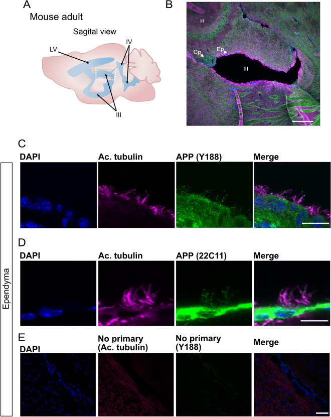
APP is localized to the ependymal cilia in adult mouse. (A) Schematic representation of adult mouse brain ventricular system, sagittal view. (B) Overview of sagittal section from adult mouse brain and the third ventricle (see dotted square in (A)) for cell nuclei stained with DAPI (blue), acetylated tubulin (magenta), APP (green). (CD) Close-up of ependymal cells and their cilia tufts (magenta) and APP expression with anti-APP Y188 antibody (C) and 22C11 antibody (D). (E) Negative controls of immunofluorescence staining without primary antibodies and only with the corresponding secondary antibodies. For cell nuclei with DAPI (blue). LV lateral ventricle, III third ventricle, IV fourth ventricle, H hippocampus, Cp choroid plexus, Ep ependyma. Magnification: (B) = 10×, (E) = 40×, (C,D) = 60×. Scale bar: (B) = 200 µm, (C,D) = 10 µm, (E) = 50 µm.

Acknowledgments
This image is the copyrighted work of the attributed author or publisher, and ZFIN has permission only to display this image to its users. Additional permissions should be obtained from the applicable author or publisher of the image. Full text @ Sci. Rep.