
|
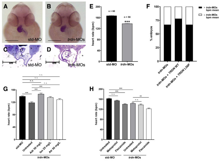
Figure 4
Heart morphology and function evaluation. (A,B) Frontal view of 48 hpf std-MO and trdn-MOs embryos hybridized with cmlc2 probe using WISH technique. Scale bars indicate 100 μm. (C,D) Semithin section of std-MO and trdn-MOs embryos in the heart region. Scale bars indicate 100 μm. (E) Quantification of heartrate in embryos at 48 hpf injected with std-MO and trdn-MOs. (F) Percentage of embryos with a heartbeat superior to the mean of the heartbeat (bpm) of trdn-MOs. (G) Heartbeat count in 3 dpf std-MO and trdn-MOs untreated or adrenaline-, isoprenaline- and atropine-treated embryos. (H) Heartbeat count in 3 dpf std-MO and trdn-MOs untreated or treated with metoprolol or flecainide. (F–H) At least 30 embryos were analyzed for each group. Values are expressed as mean ± SEM. *** p < 0.001, ** p < 0.01, n.s. = not significant, Mann–Whitney test (E) and one-way ANOVA followed by Tukey post-hoc correction (G,H). Adr, adrenaline; Iso, isoprenaline; Atr, atropine.