IMAGE

Figure 8

ID
ZDB-IMAGE-211002-11
Source
Figures for Chebli et al., 2021
Image
Figure Caption

Figure 8

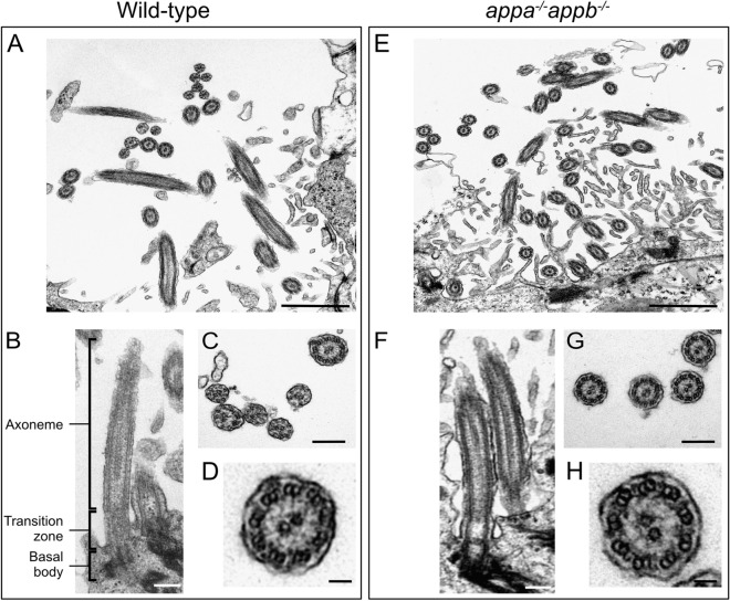
Structural integrity of ependymal cilia in WT and appa−/−appb−/− zebrafish. Transmission electron microscopy of adult zebrafish ependymal cilia of WT (AD) and appa−/−appb−/− mutant (EH) adult zebrafish. (A,E) Overview of ependymal cilia of the central canal. (B,F) Longitudinal view on the axoneme of the cilia composing its core, the transition zone including the ciliary pit between the cilia core and the cellular membrane and the basal body containing the cilia centrioles, highlighted with increased signal. In (C,G), cross-sections of cilia. (DH) Zoom on cross-section of individual cilia showing (9 + 2) microtubule doublet organization. Scale bar: (A,E) = 1 µm, (B,C,F,G) = 200 nm, (D,H) = 50 nm.

Figure Data
Acknowledgments
This image is the copyrighted work of the attributed author or publisher, and ZFIN has permission only to display this image to its users. Additional permissions should be obtained from the applicable author or publisher of the image. Full text @ Sci. Rep.