
Fig 8
- ID
- ZDB-IMAGE-210823-18
- Publication
- Waldmann et al., 2021 - The broad role of Nkx3.2 in the development of the zebrafish axial skeleton
- All Figures
- Figures for Waldmann et al., 2021
|
Fig 8
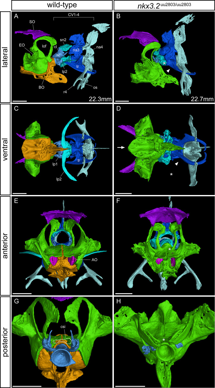
Lateral, ventral, anterior, and posterior views of wild-type (A, C, E, G) and homozygous mutant (B, D, F, H) occiput and Weberian apparatus. Cervical vertebrae (CV) 2–4 have been removed in (G, H) for clarity. Asterisk in B, D indicates the absence or severe reduction of lateral process 2 from CV2. Arrowhead in B, D indicates the absence of anterior ramus of tripus on CV3. Arrow in D indicates the V-shaped anteroventral edge of basioccipital. Asterisk in F indicates the posterior fusion of the medial gap between the paired exoccipital bones. Dotted line in G highlights the cavum sinus impar (csi), while the asterisk in H indicates its absence. The measurements in mm refer to standard length (SL). AO–asteriscus otolith, BO–basioccipital, csi—cavum sinus impar, cl–claustrum, EO–exoccipital, lof–lateral occipital fenestrae, lp–lateral process, na–neural arch, os–os suspensorium, r4 –rib 4, sc–scaphium, sn2 –supraneural 2, SO–supraoccipital, tr–tripus. Scale bars: 500μm.