IMAGE

Fig. 1

ID
ZDB-IMAGE-210814-25
Genes
Antibodies
Source
Figures for Cacialli et al., 2021
Image
Figure Caption

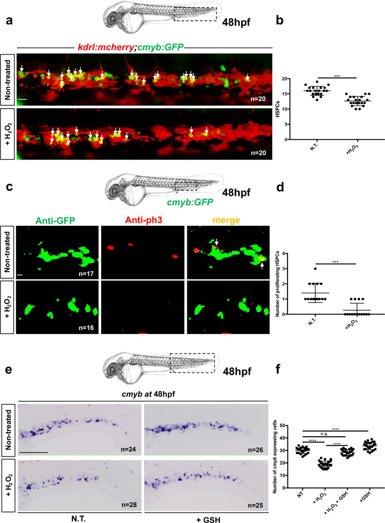
Fig. 1 Oxidative stress induces a defect in HSPC proliferation in the CHT niche at 48 hpf.

a Confocal imaging in the CHT of 48 hpf kdrl:mCherry/cmyb:GFP embryos. Embryos non-treated (NT) or incubated with H2O2 (3 mM). b Quantification of HSPCs associated with ECs. Statistical analysis: unpaired two-tailed t test, ***P < 0.001. Center values denote the mean, and error values denote s.e.m. c Anti-GFP and pH3 immunostaining of NT or H2O2-treated (3 mM) cmyb:GFP embryos. d Quantification of proliferating HSPCs; statistical analysis: unpaired two-tailed t test, ***P < 0.001. Center values denote the mean, and error values denote s.e.m. ecmyb expression at 48 hpf in NT embryos, and embryos treated with H2O2 (3 mM) or GSH (10 µm). f Quantification of cmyb-expressing cells in the CHT at 48 hpf. Statistical analysis was performed using a one-way ANOVA with Tukey–Kramer post hoc tests, adjusted for multiple comparison, ****P < 0.0001; (n.s.) non-significant P = 0.15. Center values denote the mean, and error values denote s.e.m. Scale bars: 50 μm (a); 25 μm (c); 100 μm (e).

Figure Data
Acknowledgments
This image is the copyrighted work of the attributed author or publisher, and ZFIN has permission only to display this image to its users. Additional permissions should be obtained from the applicable author or publisher of the image. Full text @ Nat. Commun.