
|
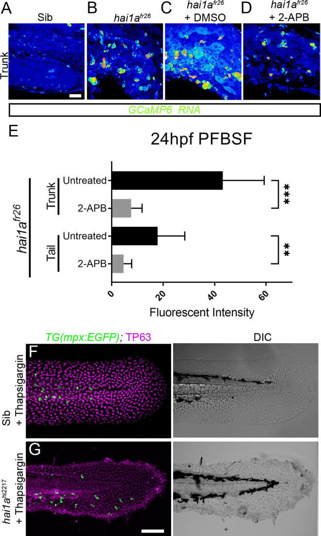
Figure 3—figure supplement 1. (A–D) Projected confocal images of eGFP in the trunk of WT (A) or hai1afr26; (B–D) injected with GCaMP6s RNA, imaged at 24hpf, indicating calcium dynamics. Embryos are either untreated (A, B), treated with DMSO (C), or with 2.5 µM 2-APB (D). Images are temporal projections of timelapse movies taken at maximum speed intervals (3 min) and projected by time. (E) Plot of pentafluorobenzenesulfonyl fluorescein (PFBSF) fluorescent staining intensity of hai1afr26 mutants at 24hpf in trunk and tail, either untreated or treated with 2-APB. n = 14; ANOVA with Bonferroni post-test ***p<0.001, **p<0.01. (F, G) Projected confocal images of tail fins of 48hpf larvae immunostained with TP63 (magenta) and eGFP (green) (left column) with DIC imaging (right column). Larvae were both hemizygous for Tg(mpx:eGFP)i114 transgene and were treated with 6.5 µM thapsigargin. Genotypes are hai1a+/hi2217 sibling (F) and hai1ahi2217 mutant (G). Rescue of neutrophil inflammation, but not epidermal defect, is apparent in treated hai1a mutant. Scale bars: (A) = 50 µm; (G) = 100 µm.