
Image
|
Figure Caption
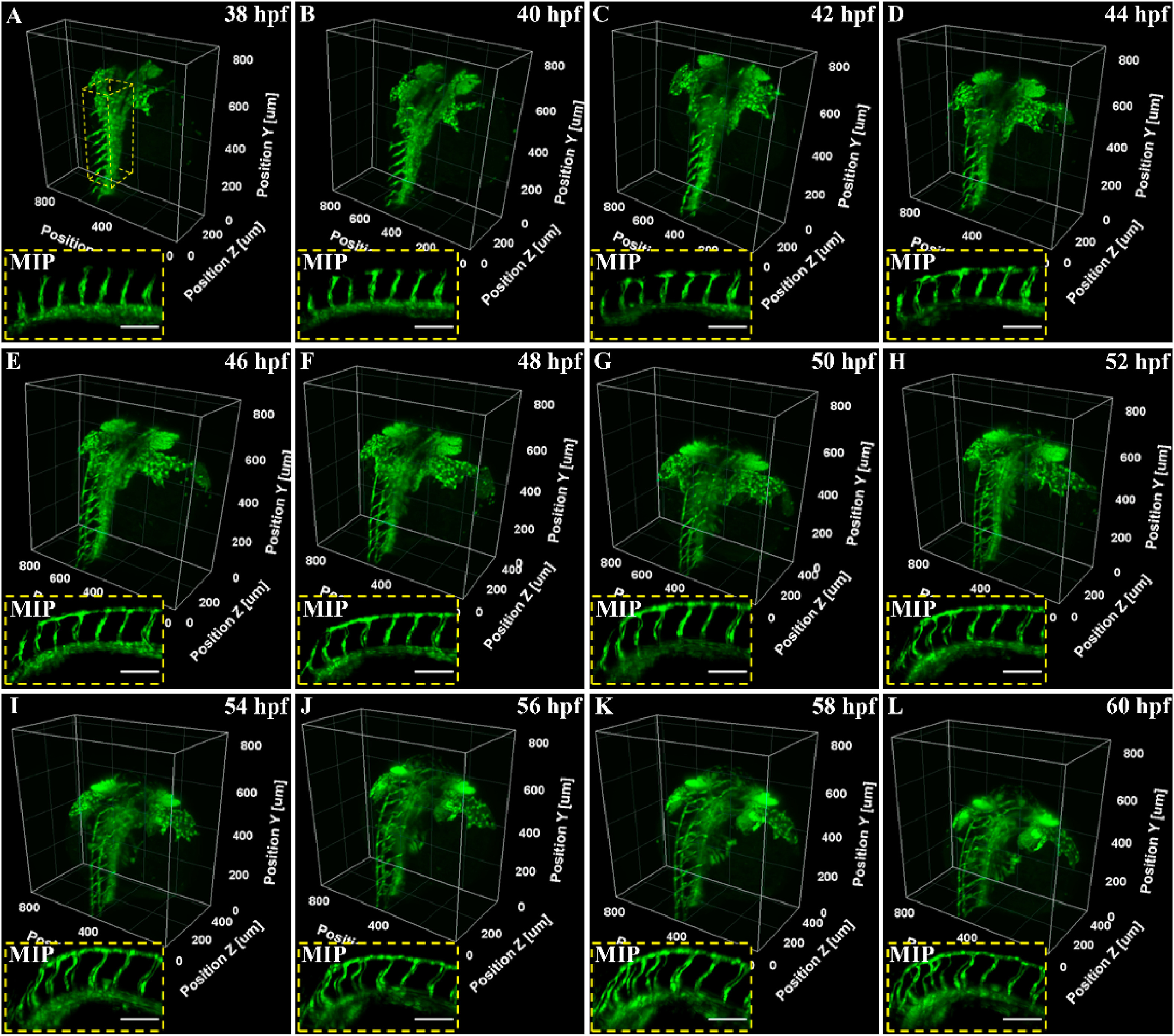
Fig. 2 3D light-sheet imaging of vascular structure in a Zebrafish trunk from 38–60 hpf with a time interval of 2 h. Insets show the corresponding 2D MIP images. Scale bar: 100 μm.
Acknowledgments
This image is the copyrighted work of the attributed author or publisher, and
ZFIN has permission only to display this image to its users.
Additional permissions should be obtained from the applicable author or publisher of the image.
Full text @ Biochem. Biophys. Res. Commun.