
Image
|
Figure Caption
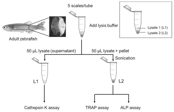
Figure 5 The schematic diagram of the scale lysis method. Five fish scales were pooled in a microcentrifuge tube. When starting an experiment, the scales were incubated in lysis buffer and then centrifuged at 17,400× g. The first half of the lysate (supernatant) was collected for cathepsin K assay. The remaining lysate was sonicated and then used for TRAP and ALP assays.
Acknowledgments
This image is the copyrighted work of the attributed author or publisher, and
ZFIN has permission only to display this image to its users.
Additional permissions should be obtained from the applicable author or publisher of the image.
Full text @ Pharmaceuticals (Basel)