
Image
|
Figure Caption
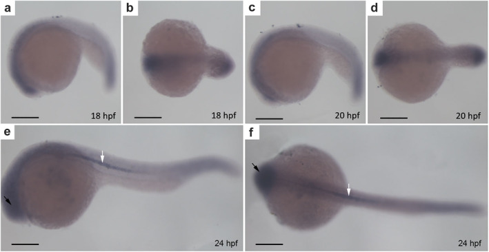
FIGURE 2
CYLD mainly localizes to the brain and notochord. (a–f) Whole‐mount in situ hybridization of zebrafish embryos. (a–b), (c–d), (e–f) side and top views of zebrafish embryos at 18 hpf, 20 hpf, and 24 hpf, respectively. The black arrows represent the brain of the zebrafish. The white arrows represent the notochord of the zebrafish. Scale bar = 250 μm
Figure Data
Acknowledgments
This image is the copyrighted work of the attributed author or publisher, and
ZFIN has permission only to display this image to its users.
Additional permissions should be obtained from the applicable author or publisher of the image.
Full text @ Thorac Cancer