IMAGE

Figure 1.

ID
ZDB-IMAGE-210611-81
Source
Figures for Koyama et al., 2021
Image
Figure Caption

Figure 1.

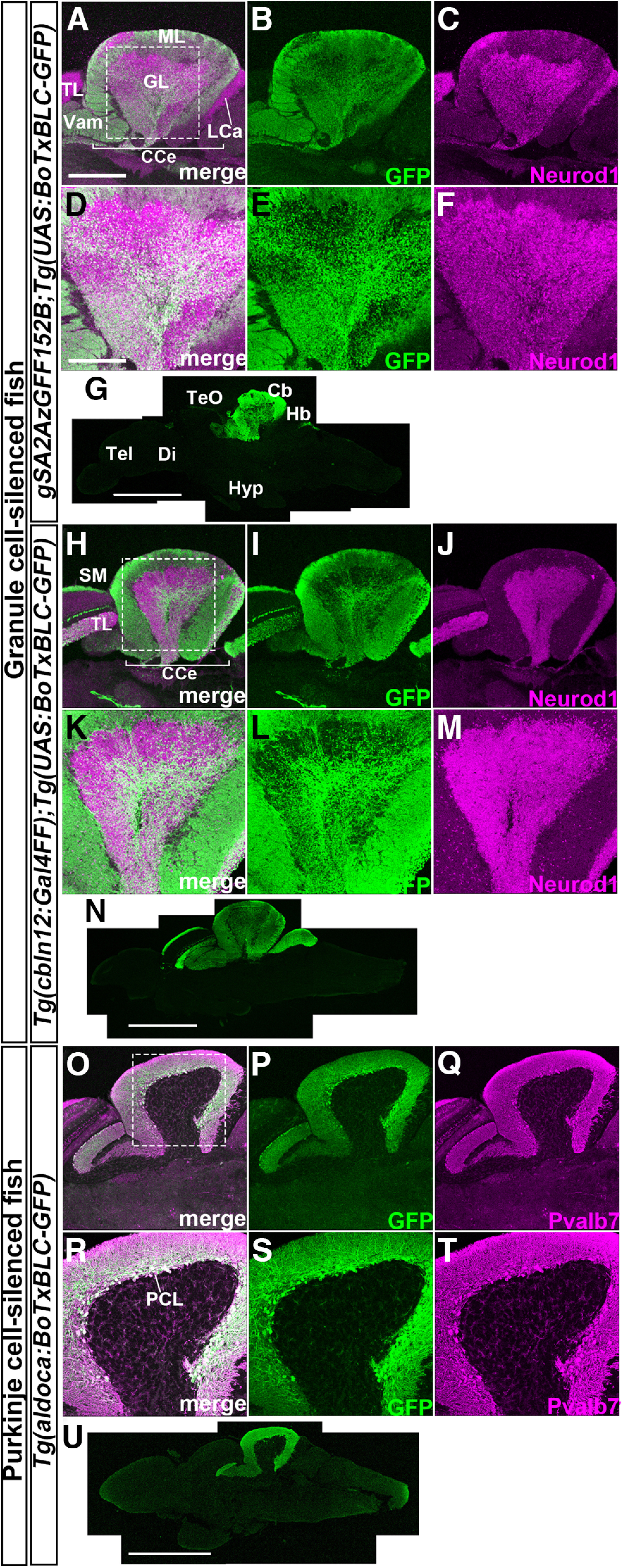
Establishment of Tg fish that express botulinum toxin in GCs or PCs. Sagittal sections of adult gSA2AzGFF152B;Tg(UAS:BoTxBLC-GFP) (A–G), Tg(cbln12:Gal4FF);Tg(UAS:BoTxBLC-GFP) (H–N), and Tg(aldoca:BoTxBLC-GFP) (O–U) brains were stained with anti-GFP (green), and anti-Neurod1, or anti-parvalbumin 7 (Pvalb7, magenta) antibodies. A–C, H–J, O–Q, Cerebellum region. D–F, K–M, R–T, High-magnification views of the boxes in A, H, O. G, N, U, Low-magnification views. Cb, cerebellum; CCe, corpus cerebelli; Di, diencephalon; GL, granular layer; Hb, hindbrain; Hyp, hypothalamus; LCa, lobus caudalis cerebelli; ML, molecular layer; PCL; PC layer; SM, stratum marginale; Tel, telencephalon; TeO, tectum opticum; TL, torus longitudinalis; Vam, medial division of valvula cerebelli. Scale bars: 400 μm (A; applies to A–C, H–J, O–Q), 200 μm (D; applies to D–F, K–M, R–T), 1 mm (G, N, U).

Acknowledgments
This image is the copyrighted work of the attributed author or publisher, and ZFIN has permission only to display this image to its users. Additional permissions should be obtained from the applicable author or publisher of the image. Full text @ eNeuro