
|
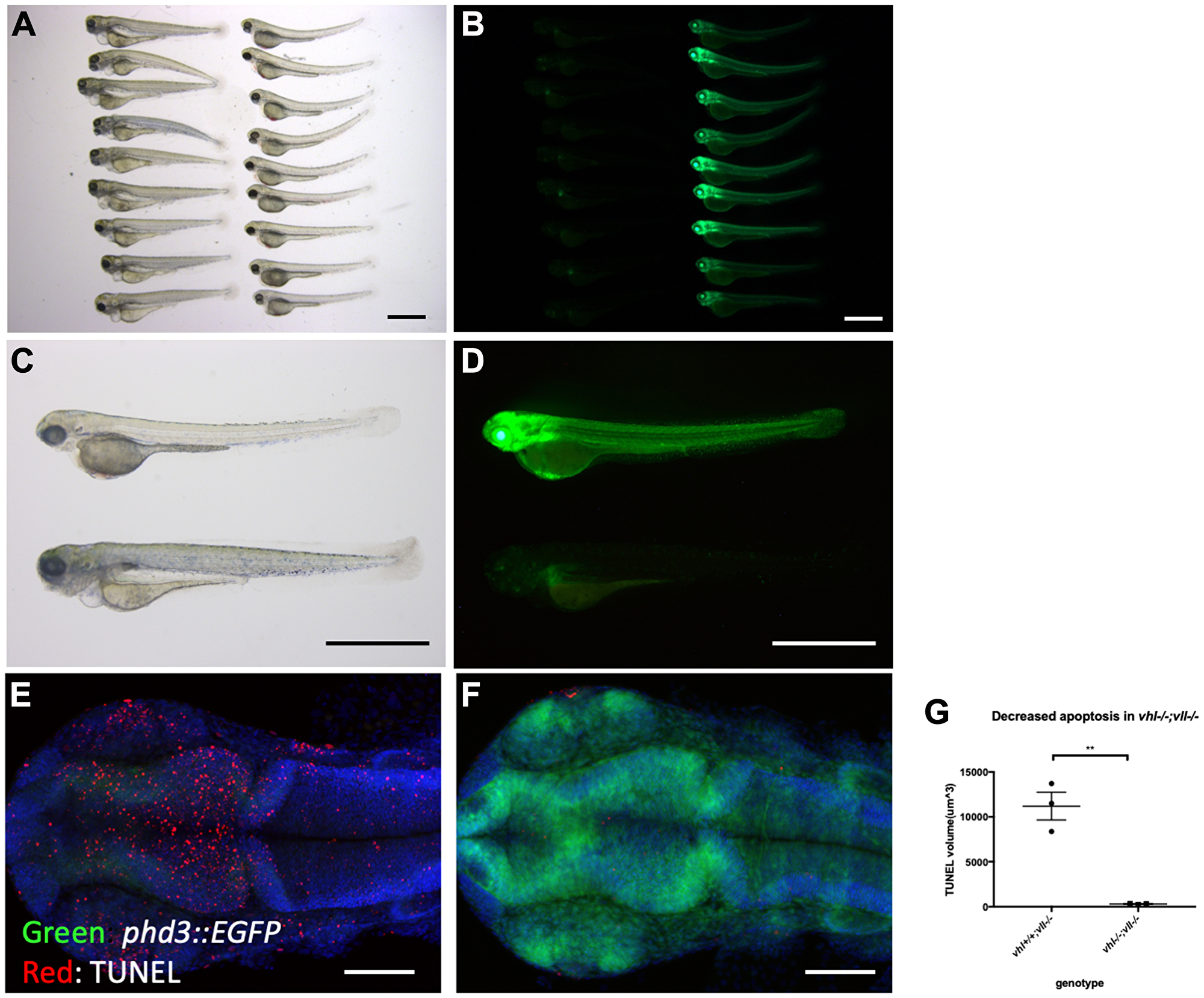
Fig. 4 vhl–/–; vll–/– double mutants are protected from X-ray induced apoptosis. (A–D) The embryos from vhl+/–; vll–/– pair mating were collected and the embryos were exposed to X-ray treatment at 1dpf. Then the treated embryos were examined at 5dpf. There were clear differences in response to X-ray treatment; majority of embryos were severely affected by X-ray treatment whereas around 25% of the embryos were well protected without much sign of cell death. We classified these embryos and looked at them under the fluorescent microscope. This revealed that all the protected embryos were EGFP positive vhl–/–; vll–/– double mutants. (E and F) The embryos were collected from the vhl+/–; vll–/– pair mating, and then exposed to X-ray at 1dpf. The embryos were fixed 3 hours post X-ray and we analysed the cell death in the head region with TUNEL. This showed extremely decreased TUNEL positive cells in the vhl–/–; vll–/– double mutants in comparison to their siblings. (G) Quantification of TUNEL stain. n = 3, **p < 0.01, unpaired t-test. Scale bars: 1 mm (A–D) and 0.2 mm (E&F).