
|
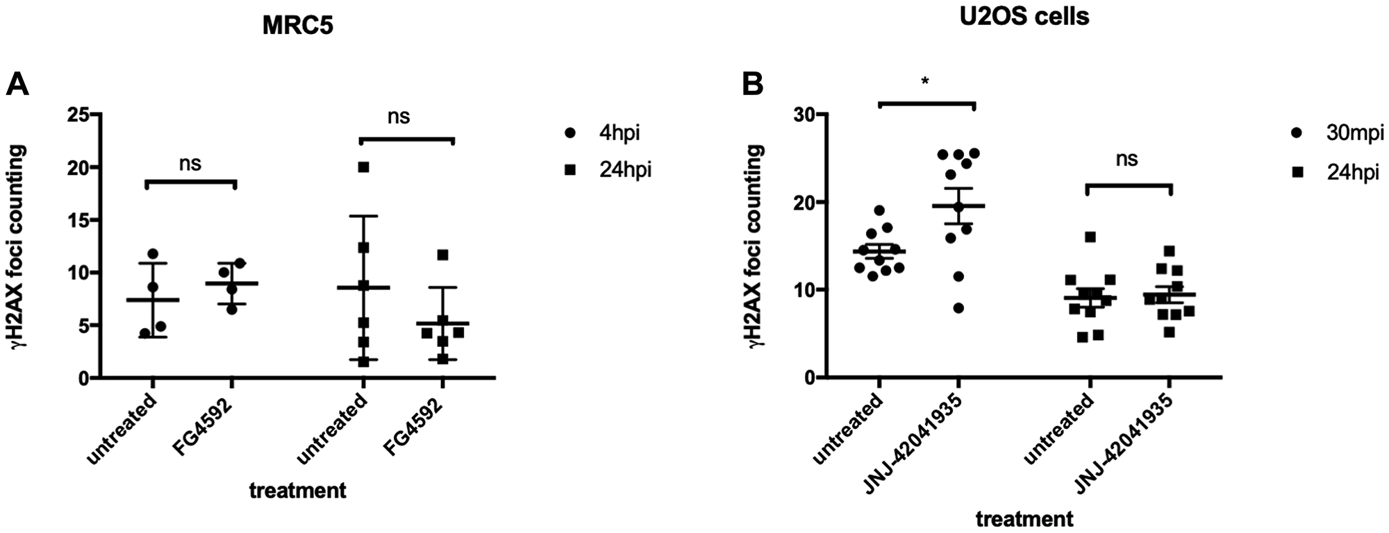
Fig. 12 HIF activation does not protect mammalian cells from X-ray induced DNA damage. (A) We treated MRC5 cells with HIF activator FG 4592 for 3 hours prior to X-ray treatment at 5 gray. The cells were fixed at 4 hpi and 24 hpi with 4% PFA and stained with γH2AX. This revealed that there was no significant difference in the γH2AX formation between HIF activator treated cells and non-treated cells. nsp > 0.05, one way ANOVA. (B) The cells were treated with HIF activator JNJ-42041935 for 3 hours prior to X-ray treatment at 2 gray. The cells were fixed at 30 mpi and 24 hpi and stained with γH2AX. At both time points, we could not observe the reduction in γH2AX formation in the HIF activator treated group in comparison to untreated group. At 30 mpi, there was even an increase in the number of γH2AX foci in the HIF activator treated group in opposition to our expectation. nsp > 0.05, *p < 0.05, one way ANOVA.