
|
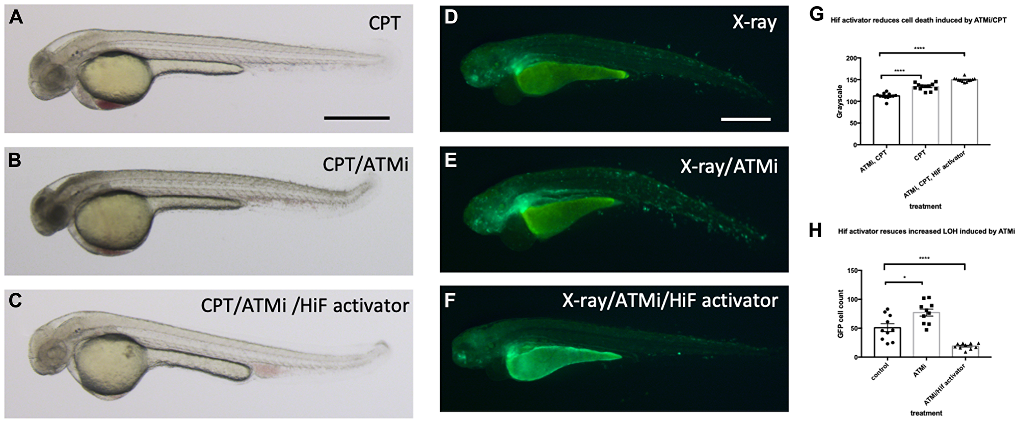
Fig. 11 Hif activation abolishes the sensitivity to CPT induced cell death in ATM inhibitor treated embryos. (A) The wild type embryos were treated with 10 nM CPT to induce cell death in the CNS. (B) When the embryos were treated with ATMi before CPT treatment, the embryos were highly sensitised to CPT treatment and induced severe cell death in the CNS. (C) When the embryos were treated with Hif activator in combination with ATMi prior to CPT treatment, the embryos were well protected from CPT/ATMi induced cell death in CNS. The quantification of the data is shown in (G) ****p < 0.0001, one way ANOVA. (D) The vhl+/–; vll–/–; Tg (phd3:: EGFP)i144 embryos were treated with X-ray to induce LOH at the vhl locus. (E) When the vhl+/–; vll–/–; Tg (phd3:: EGFP)i144 embryos were treated with ATMi before the X-ray treatment, there was a dramatic increase in the LOH in the vhl locus in the ATMi treated embryos. (F) When the embryos were treated with Hif activator in combination with ATMi before the X-ray treatment, there was a remarkable reduction in the LOH in the vhl locus. The quantification of the data is shown in (H) *p < 0.05, ****p < 0.0001, one way ANOVA. Scale bars: 0.5 mm.