
|
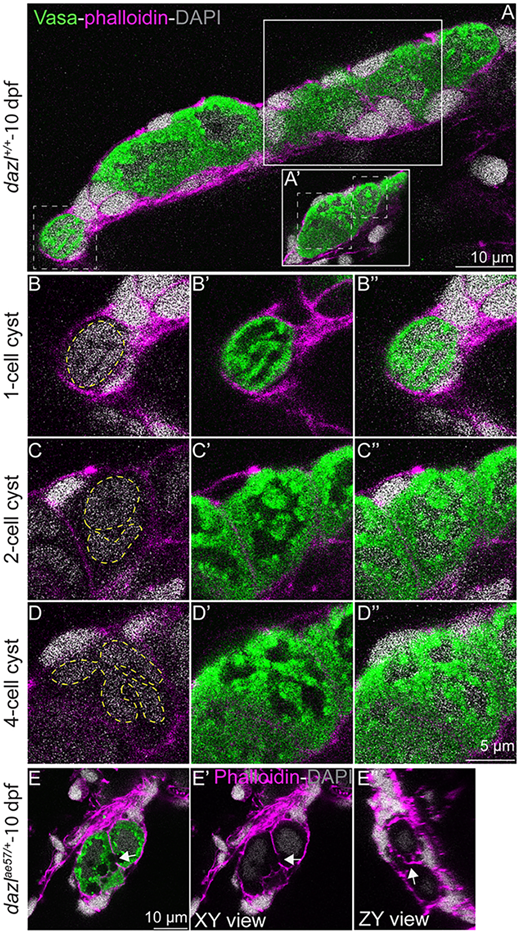
Fig. 1 (A,A′) Single confocal plane of a 10-day larval gonad with DAPI-labeled nuclei (gray), GCs marked with Vasa (green), and F-actin with phalloidin (pink). Cystogenesis stages are boxed (white dotted lines). Solid white boxes delineate the focal plane showing the two- or four-cell stage cysts. (B-B″) Images showing a one-cell stage cyst with compact DNA (B) and a high cytoplasm/nucleus ratio (B′). Yellow dashed line indicates the nucleus. (C-C″) Division produces a two-cell stage cyst with two nuclei (yellow dashed line) surrounded by perinuclear Vasa. (D-D″) A four-cell cyst with four nuclei (yellow dashed line). (E-E″) Intercellular bridges (white arrow) in a 10-day larval gonad. (E′) XY view without Vasa (green). White arrow indicates the intercellular bridge. (E″) ZY view of the actin ring (white arrow).