
|
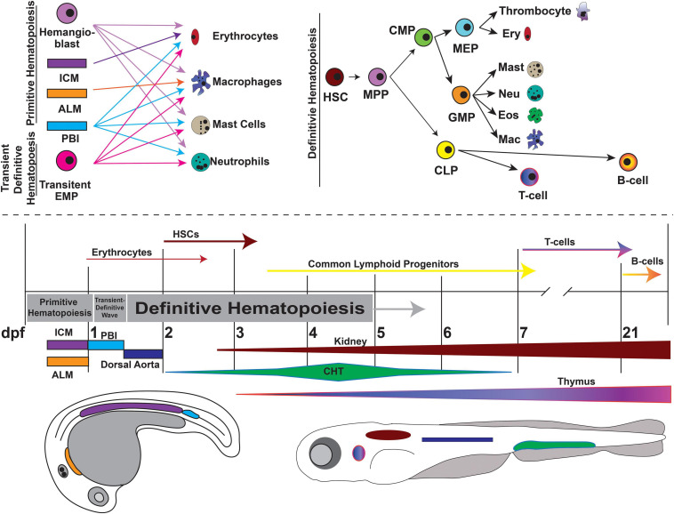
FIGURE 5
Overview of zebrafish hematopoiesis. Zebrafish hematopoiesis occurs in two waves, primitive and definitive, the latter of which recapitulates all the same blood cell types as humans. During the primitive wave, hemangioblasts give rise to a portion of primitive erythrocytes and granulocytes. The main source of primitive granulocyte production occurs within the anterior lateral mesoderm (ALM) (orange), while primitive erythrocyte productions occurs in the intermediate cell mass (ICM) (purple). Once circulation begins at approximately 24 hpf, primitive blood production transitions to the posterior blood island (PBI) (blue). Between 24 and 36 hpf a transient wave occurs in which transient bipotential erythromyeloid progenitor cells (EMPs). By 36 hpf, the first hematopoietic stem cells (HSCs) arise from the ventral wall of the dorsal aorta (navy), a site analogous to the aorta-gonad-mesonephros (AGM) in mammals. Once HSCs arise, they migrate to the caudal hematopoietic tissue (CHT) (previously the PBI), a structure analogous to the mammalian fetal liver. It is in the CHT that definitive erythroid and myeloid cells are produced and replace their primitive counterparts. By 4dpf HSCs migrate to and seed the kidney marrow (dark red), the equivalent of bone marrow in mammals. The first T-cells originate at approximately 7 dpf and mature in the thymus (purple/pink), while B-cells are not present until 21 dpf. This figure was constructed using information from published reviews (