IMAGE

FIGURE 3

ID
ZDB-IMAGE-210518-53
Source
Figures for Wiweger et al., 2021
Image
Figure Caption

FIGURE 3

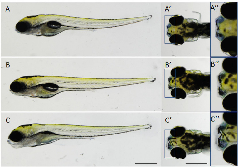
Specificity of Nile blue staining in the olfactory organ in npc2–/– larvae at 5 dpf. (A–A”) untreated npc2–/– larvae; (B–B”) treatment with 2 mM 2-hydroxypropyl-β-cyclodextrin (2HPβCD; a drug that is used in clinical trials to treat NPC patients) decreased staining intensity to wildtype levels (Figures 2A–A”); and (C–C”) treatment with 8 mM (3β)-3-(2-[diethylamino]ethoxy)androst-5-en-17-one hydrochloride (U18666A, an inhibitor of cholesterol synthesis that is used to model NPC chemically) increased staining intensity in npc2–/– larvae. (A–C) lateral view; (A’,A”,B’,B”,C’,C”), dorsal view; and (A”,B”,C”) enlarged fragments of A’,B’,C’, respectively. Scale bar = 0.5 mm.

Figure Data
Acknowledgments
This image is the copyrighted work of the attributed author or publisher, and ZFIN has permission only to display this image to its users. Additional permissions should be obtained from the applicable author or publisher of the image. Full text @ Front. Cell. Neurosci.