
|
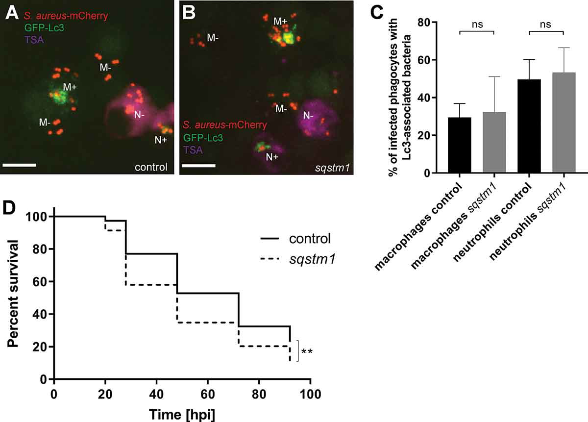
Fig. 9 Loss of Sqstm1 leads to increased susceptibility to S. aureus infection. (A and B) Confocal photomicrographs are shown as maximum intensity projections of the Lc3-mediated response at 1 hpi within infected macrophages and neutrophils of control (A) and sqstm1 knockdown (B) fixed CMV:GFP-Lc3 embryos infected with mCherry-labeled S. aureus. Embryos were fixed at 1 hpi and chemically stained for Mpx activity (TSA, magenta). TSA-negative macrophages are seen containing bacteria with (M+) or without (M-) Lc3 aggregates. TSA-positive neutrophils contain bacteria with (N+) and without Lc3 aggregates (N-). The images shown are representative of three independent experiments. Scale bars:10 µm. (C) Quantification of Lc3 associations with intracellular S. aureus at 1 hpi within infected macrophages and neutrophils of control and sqstm1 knockdown fixed CMV:GFP-Lc3 embryos. Data are shown as mean ± standard deviation (SD) obtained from three independent experiments. 174 infected macrophages and 72 neutrophils were analyzed in 18 control larvae. 165 infected macrophages and 68 neutrophils were analyzed in sqstm1 knockdown larvae. One-way ANOVA with Bonferroni’s posttest was used. ns – not significant. (D) Survival of irf8-only or irf8 + sqstm1 knockdown zebrafish larvae following intravenous injection with S. aureus at 30 hpf (≥69 larvae per group). This result is obtained from three independent experiments. Survival curves were compared using a log-rank (Mantel-Cox) statistical test. ** P < 0.01