
|
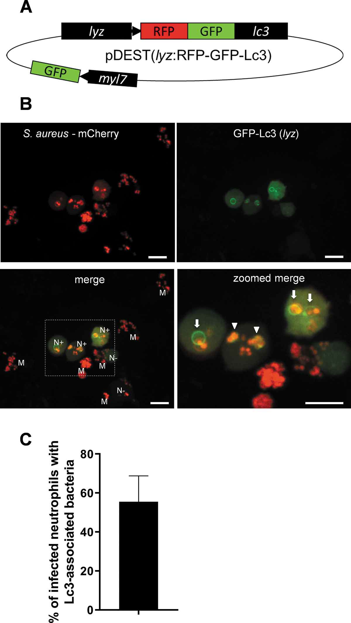
Fig. 2 Generation of a lyz:RFP-GFP-Lc3 transgenic line in zebrafish confirms the Lc3-mediated response to S. aureus within neutrophils. (A) Schematic of the pDEST(lyz:RFP-GFP-Lc3) construct encoding the fusion RFP-GFP-Lc3 protein under the neutrophil-specific lyz promoter. In addition, the heart marker myl7-driven GFP is used to facilitate the screening of positive larvae. (B) Confocal images at maximum projection of the Lc3-mediated response at 1 hpi in live lyz:RFP-GFP-Lc3 embryos infected with approximately 1500 CFU of mCherry-labeled S. aureus. Lyz-positive neutrophils are seen containing bacteria with (N+) or without (N-) Lc3 aggregates. Lyz-negative macrophages are also seen containing bacteria (M). The images shown are representative of three independent experiments. Arrows indicate spacious Lc3-positive vesicles, whereas arrowheads show tightly wrapped Lc3-associated bacteria. Scale bars: 10 µm. (C) Quantification of Lc3 associations with intracellular S. aureus within infected neutrophils of live lyz:RFP-GFP-Lc3 embryos at 1 hpi with approximately 1500 CFU. Data are shown as mean ± standard deviation (SD) obtained from three independent experiments (6 larvae per experiment). 79 infected neutrophils were analyzed in 18 larvae