
Image
|
Figure Caption
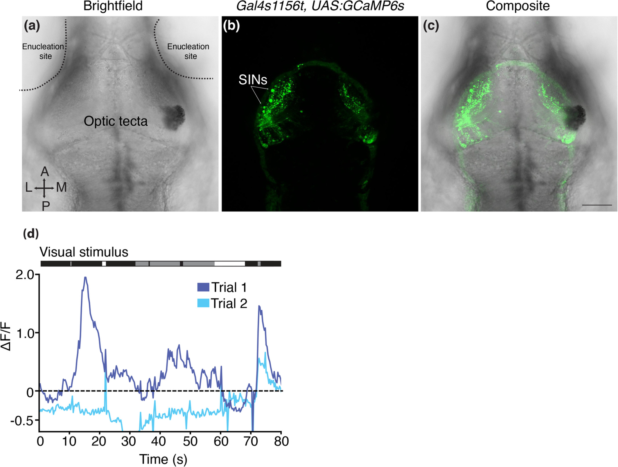
Fig. 5 Gal4s1156t+ SIN whole‐field luminance responses require the retina. (a–c) 7 dpf Gal4s1156t × UAS:GCaMP6s larva with bilateral removal of retinal input. Enucleation sites and optic tecta are denoted in the brightfield image (a). Following enucleation, GCaMP6s+ SIN cell bodies are still clearly visible (b, c). (d) Example SIN from the larva shown in (a–c). Responses to luminance transitions are not consistent across trials and are not closely synced to the visual stimuli (shown above trace). Scale bar (a–c) = 100 μm
Acknowledgments
This image is the copyrighted work of the attributed author or publisher, and
ZFIN has permission only to display this image to its users.
Additional permissions should be obtained from the applicable author or publisher of the image.
Full text @ J. Comp. Neurol.