
Image
|
Figure Caption
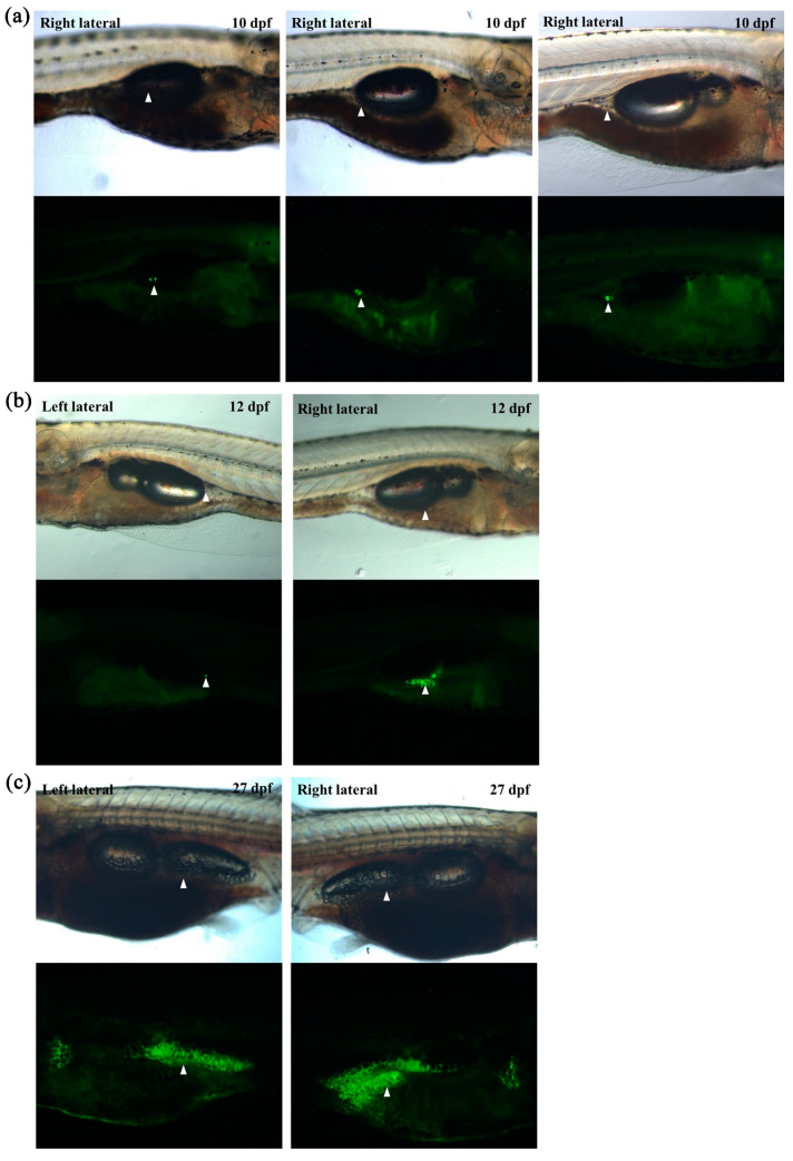
Figure 3 In vivo monitoring of visceral adipose tissue development in larvae using transgenic zebrafish. (a) Representative images of the first adipocyte emerging in three independent larvae at 10 dpf. (b) Representative images of visceral adipose tissues at 12 dpf. The first adipocyte of left lateral emerged. (c) Representative images of visceral adipose tissues at 27 dpf. White arrowheads indicate visceral adipose tissue.
Acknowledgments
This image is the copyrighted work of the attributed author or publisher, and
ZFIN has permission only to display this image to its users.
Additional permissions should be obtained from the applicable author or publisher of the image.
Full text @ Int. J. Mol. Sci.